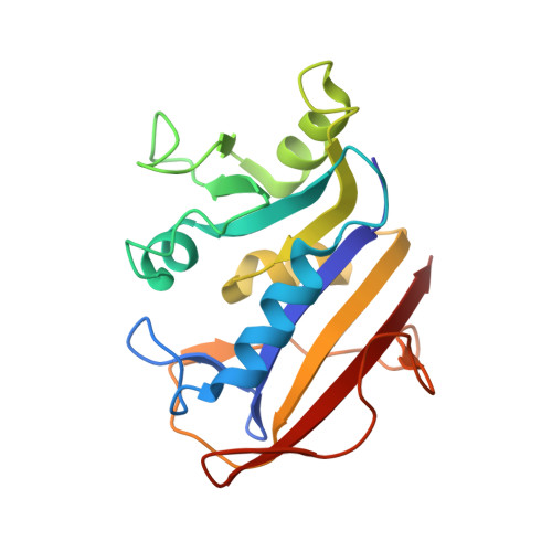
Preferential selection of isomer binding from chiral mixtures: alternate binding modes observed for the E and Z isomers of a series of 5-substituted 2,4-diaminofuro[2,3-d]pyrimidines as ternary complexes with NADPH and human dihydrofolate reductase.
Cody, V., Piraino, J., Pace, J., Li, W., Gangjee, A.(2010) Acta Crystallogr D Biol Crystallogr 66: 1271-1277
- PubMed: 21123866
- DOI: https://doi.org/10.1107/S0907444910035808
- Primary Citation of Related Structures:
3NXO, 3NXR, 3NXT, 3NXV, 3NXX, 3NXY - PubMed Abstract:
The crystal structures of six human dihydrofolate reductase (hDHFR) ternary complexes with NADPH and a series of mixed E/Z isomers of 5-substituted 5-[2-(2-methoxyphenyl)-prop-1-en-1-yl]furo[2,3-d]pyrimidine-2,4-diamines substituted at the C9 position with propyl, isopropyl, cyclopropyl, butyl, isobutyl and sec-butyl (E2-E7, Z3) were determined and the results were compared with the resolved E and Z isomers of the C9-methyl parent compound. The configuration of all of the inhibitors, save one, was observed as the E isomer, in which the binding of the furopyrimidine ring is flipped such that the 4-amino group binds in the 4-oxo site of folate. The Z3 isomer of the C9-isopropyl analog has the normal 2,4-diaminopyrimidine ring binding geometry, with the furo oxygen near Glu30 and the 4-amino group interacting near the cofactor nicotinamide ring. Electron-density maps for these structures revealed the binding of only one isomer to hDHFR, despite the fact that chiral mixtures (E:Z ratios of 2:1, 3:1 and 3:2) of the inhibitors were incubated with hDHFR prior to crystallization. Superposition of the hDHFR complexes with E2 and Z3 shows that the 2'-methoxyphenyl ring of E2 is perpendicular to that of Z3. The most potent inhibitor in this series is the isopropyl analog Z3 and the least potent is the isobutyl analog E6, consistent with data that show that the Z isomer makes the most favorable interactions with the active-site residues. The isobutyl moiety of E6 is observed in two orientations and the resultant steric crowding of the E6 analog is consistent with its weaker activity. The alternative binding modes observed for the furopyrimidine ring in these E/Z isomers suggest that new templates can be designed to probe these binding regions of the DHFR active site.
- Structural Biology Department, Hauptman-Woodward Medical Research Institute, 700 Ellicott Street, Buffalo, NY 14203, USA. cody@hwi.buffalo.edu
Organizational Affiliation:


