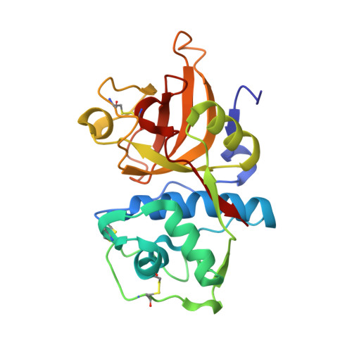
3OF8

Structural Basis for Reversible and Irreversible Inhibition of Human Cathepsin L by their Respective Dipeptidyl Glyoxal and Diazomethylketone Inhibitors


Experimental Data Snapshot

  • Method: X-RAY DIFFRACTION
  • Resolution: 2.20 Å
  • R-Value Free: 0.253 
  • R-Value Work: 0.194 
  • R-Value Observed: 0.197 

Starting Model: experimental
View more details

wwPDB Validation   3D Report Full Report


Ligand Structure Quality Assessment 


This is version 1.6 of the entry. See complete history


Literature

Structural basis for reversible and irreversible inhibition of human cathepsin L by their respective dipeptidyl glyoxal and diazomethylketone inhibitors.

Shenoy, R.T.Sivaraman, J.

(2011) J Struct Biol 173: 14-19

  • DOI: https://doi.org/10.1016/j.jsb.2010.09.007
  • Primary Citation of Related Structures:  
    3OF8, 3OF9

  • PubMed Abstract: 

    Cathepsin L plays a key role in many pathophysiological conditions including rheumatoid arthritis, tumor invasion and metastasis, bone resorption and remodeling. Here we report the crystal structures of two analogous dipeptidyl inhibitor complexes which inhibit human cathepsin L in reversible and irreversible modes, respectively. To-date, there are no crystal structure reports of complexes of proteases with their glyoxal inhibitors or complexes of cathepsin L and their diazomethylketone inhibitors. These two inhibitors - inhibitor 1, an α-keto-β-aldehyde and inhibitor 2, a diazomethylketone, have different groups in the S1 subsite. Inhibitor 1 [Z-Phe-Tyr (OBut)-COCHO], with a K(i) of 0.6nM, is the most potent, reversible, synthetic peptidyl inhibitor of cathepsin L reported to-date. The structure of the inhibitor 1 complex was refined up to 2.2Å resolution. The structure of the complex of the inhibitor 2 [Z-Phe-Tyr (t-Bu)-diazomethylketone], an irreversible inhibitor that can inactivate cathepsin L at μM concentrations, was refined up to 1.76Å resolution. These two inhibitors have substrate-like interactions with the active site cysteine (Cys25). Inhibitor 1 forms a tetrahedral hemithioacetal adduct, whereas the inhibitor 2 forms a thioester with Cys25. The inhibitor 1 β-aldehyde group is shown to make a hydrogen bond with catalytic His163, whereas the ketone carbonyl oxygen of the inhibitor 2 interacts with the oxyanion hole. tert-Butyl groups of both inhibitors are found to make several non-polar contacts with S' subsite residues of cathepsin L. These studies, combined with other complex structures of cathepsin L, reveal the structural basis for their potency and selectivity.


  • Organizational Affiliation

    Department of Biological Sciences, National University of Singapore, Singapore 117543, Singapore.


Macromolecules
Find similar proteins by:  (by identity cutoff)  |  3D Structure
Entity ID: 1
MoleculeChains Sequence LengthOrganismDetailsImage
Cathepsin L1221Homo sapiensMutation(s): 0 
Gene Names: CTSL1CTSL
EC: 3.4.22.15
UniProt & NIH Common Fund Data Resources
Find proteins for P07711 (Homo sapiens)
Explore P07711 
Go to UniProtKB:  P07711
PHAROS:  P07711
GTEx:  ENSG00000135047 
Entity Groups  
Sequence Clusters30% Identity50% Identity70% Identity90% Identity95% Identity100% Identity
UniProt GroupP07711
Sequence Annotations
Expand
  • Reference Sequence
Small Molecules
Ligands 1 Unique
IDChains Name / Formula / InChI Key2D Diagram3D Interactions
I0Y
Query on I0Y

Download Ideal Coordinates CCD File 
B [auth A]Nalpha-[(benzyloxy)carbonyl]-N-[(2S)-1-(4-tert-butoxyphenyl)-4-hydroxy-3-oxobutan-2-yl]-L-phenylalaninamide
C31 H36 N2 O6
DKORZCNOWZIKDV-SVBPBHIXSA-N
Binding Affinity Annotations 
IDSourceBinding Affinity
I0Y PDBBind:  3OF8 Ki: 0.6 (nM) from 1 assay(s)
Experimental Data & Validation

Experimental Data

  • Method: X-RAY DIFFRACTION
  • Resolution: 2.20 Å
  • R-Value Free: 0.253 
  • R-Value Work: 0.194 
  • R-Value Observed: 0.197 
  • Space Group: P 65
Unit Cell:
Length ( Å )Angle ( ˚ )
a = 85.318α = 90
b = 85.318β = 90
c = 50.018γ = 120
Software Package:
Software NamePurpose
PHENIXrefinement
SCALEPACKdata scaling
MOLREPphasing
CNSrefinement
PDB_EXTRACTdata extraction
HKL-2000data reduction
HKL-2000data scaling
DENZOdata reduction

Structure Validation

View Full Validation Report


Ligand Structure Quality Assessment 


Entry History 

Deposition Data

Revision History  (Full details and data files)

  • Version 1.0: 2010-12-08
    Type: Initial release
  • Version 1.1: 2011-07-13
    Changes: Version format compliance
  • Version 1.2: 2012-12-12
    Changes: Other
  • Version 1.3: 2017-11-08
    Changes: Advisory, Refinement description
  • Version 1.4: 2019-07-17
    Changes: Data collection, Derived calculations, Refinement description
  • Version 1.5: 2023-09-06
    Changes: Advisory, Data collection, Database references, Derived calculations, Refinement description
  • Version 1.6: 2024-10-09
    Changes: Structure summary