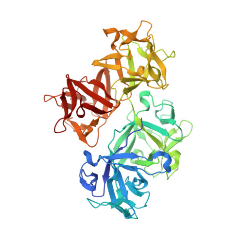
3P53

Structure of fascin


Experimental Data Snapshot

  • Method: X-RAY DIFFRACTION
  • Resolution: 2.00 Å
  • R-Value Free: 0.220 
  • R-Value Work: 0.178 
  • R-Value Observed: 0.179 

wwPDB Validation   3D Report Full Report


This is version 1.4 of the entry. See complete history


Literature

Mechanism of actin filament bundling by fascin.

Jansen, S.Collins, A.Yang, C.Rebowski, G.Svitkina, T.Dominguez, R.

(2011) J Biol Chem 286: 30087-30096

  • DOI: https://doi.org/10.1074/jbc.M111.251439
  • Primary Citation of Related Structures:  
    3P53

  • PubMed Abstract: 

    Fascin is the main actin filament bundling protein in filopodia. Because of the important role filopodia play in cell migration, fascin is emerging as a major target for cancer drug discovery. However, an understanding of the mechanism of bundle formation by fascin is critically lacking. Fascin consists of four β-trefoil domains. Here, we show that fascin contains two major actin-binding sites, coinciding with regions of high sequence conservation in β-trefoil domains 1 and 3. The site in β-trefoil-1 is located near the binding site of the fascin inhibitor macroketone and comprises residue Ser-39, whose phosphorylation by protein kinase C down-regulates actin bundling and formation of filopodia. The site in β-trefoil-3 is related by pseudo-2-fold symmetry to that in β-trefoil-1. The two sites are ∼5 nm apart, resulting in a distance between actin filaments in the bundle of ∼8.1 nm. Residue mutations in both sites disrupt bundle formation in vitro as assessed by co-sedimentation with actin and electron microscopy and severely impair formation of filopodia in cells as determined by rescue experiments in fascin-depleted cells. Mutations of other areas of the fascin surface also affect actin bundling and formation of filopodia albeit to a lesser extent, suggesting that, in addition to the two major actin-binding sites, fascin makes secondary contacts with other filaments in the bundle. In a high resolution crystal structure of fascin, molecules of glycerol and polyethylene glycol are bound in pockets located within the two major actin-binding sites. These molecules could guide the rational design of new anticancer fascin inhibitors.


  • Organizational Affiliation

    Department of Physiology, University of Pennsylvania School of Medicine, Philadelphia, Pennsylvania 19104, USA.


Macromolecules
Find similar proteins by:  (by identity cutoff)  |  3D Structure
Entity ID: 1
MoleculeChains Sequence LengthOrganismDetailsImage
Fascin
A, B
496Homo sapiensMutation(s): 0 
Gene Names: FAN1FSCN1HSNHumanSNL
UniProt & NIH Common Fund Data Resources
Find proteins for Q16658 (Homo sapiens)
Explore Q16658 
Go to UniProtKB:  Q16658
PHAROS:  Q16658
GTEx:  ENSG00000075618 
Entity Groups  
Sequence Clusters30% Identity50% Identity70% Identity90% Identity95% Identity100% Identity
UniProt GroupQ16658
Sequence Annotations
Expand
  • Reference Sequence
Small Molecules
Ligands 4 Unique
IDChains Name / Formula / InChI Key2D Diagram3D Interactions
12P
Query on 12P

Download Ideal Coordinates CCD File 
G [auth A]DODECAETHYLENE GLYCOL
C24 H50 O13
WRZXKWFJEFFURH-UHFFFAOYSA-N
1PE
Query on 1PE

Download Ideal Coordinates CCD File 
H [auth A],
I [auth A],
J [auth A],
O [auth B],
P [auth B]
PENTAETHYLENE GLYCOL
C10 H22 O6
JLFNLZLINWHATN-UHFFFAOYSA-N
PG0
Query on PG0

Download Ideal Coordinates CCD File 
Q [auth B]2-(2-METHOXYETHOXY)ETHANOL
C5 H12 O3
SBASXUCJHJRPEV-UHFFFAOYSA-N
GOL
Query on GOL

Download Ideal Coordinates CCD File 
C [auth A]
D [auth A]
E [auth A]
F [auth A]
K [auth B]
C [auth A],
D [auth A],
E [auth A],
F [auth A],
K [auth B],
L [auth B],
M [auth B],
N [auth B]
GLYCEROL
C3 H8 O3
PEDCQBHIVMGVHV-UHFFFAOYSA-N
Experimental Data & Validation

Experimental Data

  • Method: X-RAY DIFFRACTION
  • Resolution: 2.00 Å
  • R-Value Free: 0.220 
  • R-Value Work: 0.178 
  • R-Value Observed: 0.179 
  • Space Group: C 1 2 1
Unit Cell:
Length ( Å )Angle ( ˚ )
a = 161.693α = 90
b = 71.01β = 131.23
c = 112.736γ = 90
Software Package:
Software NamePurpose
HKL-2000data collection
SnBphasing
PHENIXrefinement
HKL-2000data reduction
HKL-2000data scaling

Structure Validation

View Full Validation Report


Entry History 

Deposition Data

Revision History  (Full details and data files)

  • Version 1.0: 2011-06-29
    Type: Initial release
  • Version 1.1: 2011-07-13
    Changes: Version format compliance
  • Version 1.2: 2011-09-07
    Changes: Database references
  • Version 1.3: 2014-04-16
    Changes: Other
  • Version 1.4: 2024-02-21
    Changes: Data collection, Database references, Derived calculations