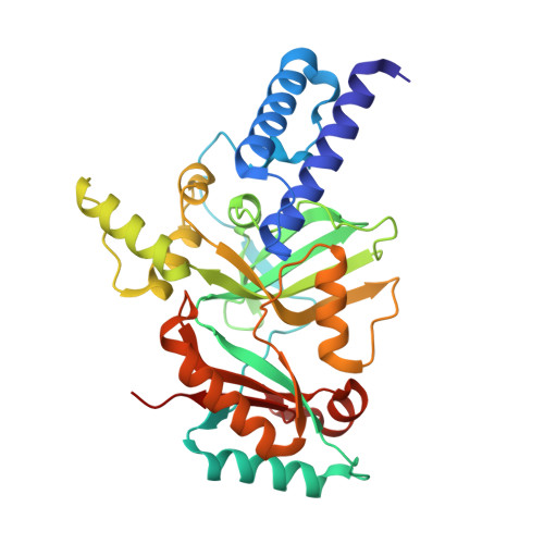
3PN1

Novel Bacterial NAD+-dependent DNA Ligase Inhibitors with Broad Spectrum Potency and Antibacterial Efficacy In Vivo


Experimental Data Snapshot

  • Method: X-RAY DIFFRACTION
  • Resolution: 2.00 Å
  • R-Value Free: 0.231 
  • R-Value Work: 0.201 
  • R-Value Observed: 0.203 

wwPDB Validation   3D Report Full Report


Ligand Structure Quality Assessment 


This is version 1.4 of the entry. See complete history


Literature

Novel Bacterial NAD+-Dependent DNA Ligase Inhibitors with Broad-Spectrum Activity and Antibacterial Efficacy In Vivo.

Mills, S.D.Eakin, A.E.Buurman, E.T.Newman, J.V.Gao, N.Huynh, H.Johnson, K.D.Lahiri, S.Shapiro, A.B.Walkup, G.K.Yang, W.Stokes, S.S.

(2011) Antimicrob Agents Chemother 55: 1088-1096

  • DOI: https://doi.org/10.1128/AAC.01181-10
  • Primary Citation of Related Structures:  
    3PN1

  • PubMed Abstract: 

    DNA ligases are indispensable enzymes playing a critical role in DNA replication, recombination, and repair in all living organisms. Bacterial NAD+-dependent DNA ligase (LigA) was evaluated for its potential as a broad-spectrum antibacterial target. A novel class of substituted adenosine analogs was discovered by target-based high-throughput screening (HTS), and these compounds were optimized to render them more effective and selective inhibitors of LigA. The adenosine analogs inhibited the LigA activities of Escherichia coli, Haemophilus influenzae, Mycoplasma pneumoniae, Streptococcus pneumoniae, and Staphylococcus aureus, with inhibitory activities in the nanomolar range. They were selective for bacterial NAD+-dependent DNA ligases, showing no inhibitory activity against ATP-dependent human DNA ligase 1 or bacteriophage T4 ligase. Enzyme kinetic measurements demonstrated that the compounds bind competitively with NAD+. X-ray crystallography demonstrated that the adenosine analogs bind in the AMP-binding pocket of the LigA adenylation domain. Antibacterial activity was observed against pathogenic Gram-positive and atypical bacteria, such as S. aureus, S. pneumoniae, Streptococcus pyogenes, and M. pneumoniae, as well as against Gram-negative pathogens, such as H. influenzae and Moraxella catarrhalis. The mode of action was verified using recombinant strains with altered LigA expression, an Okazaki fragment accumulation assay, and the isolation of resistant strains with ligA mutations. In vivo efficacy was demonstrated in a murine S. aureus thigh infection model and a murine S. pneumoniae lung infection model. Treatment with the adenosine analogs reduced the bacterial burden (expressed in CFU) in the corresponding infected organ tissue as much as 1,000-fold, thus validating LigA as a target for antibacterial therapy.


  • Organizational Affiliation

    AstraZeneca R&D Boston, 35 Gatehouse Drive, Waltham, MA 02451, USA. Scott.Mills@astrazeneca.com


Macromolecules
Find similar proteins by:  (by identity cutoff)  |  3D Structure
Entity ID: 1
MoleculeChains Sequence LengthOrganismDetailsImage
DNA ligase318Haemophilus influenzaeMutation(s): 0 
Gene Names: ligAligligNHI_1100
EC: 6.5.1.2
UniProt
Find proteins for P43813 (Haemophilus influenzae (strain ATCC 51907 / DSM 11121 / KW20 / Rd))
Explore P43813 
Go to UniProtKB:  P43813
Entity Groups  
Sequence Clusters30% Identity50% Identity70% Identity90% Identity95% Identity100% Identity
UniProt GroupP43813
Sequence Annotations
Expand
  • Reference Sequence
Small Molecules
Ligands 2 Unique
IDChains Name / Formula / InChI Key2D Diagram3D Interactions
IVH
Query on IVH

Download Ideal Coordinates CCD File 
B [auth A]2-(butylsulfanyl)adenosine
C14 H21 N5 O4 S
JUZWTKSKLZRPBL-QYVSTXNMSA-N
IWH
Query on IWH

Download Ideal Coordinates CCD File 
C [auth A]1-(2,4-dimethylbenzyl)-6-oxo-1,6-dihydropyridine-3-carboxamide
C15 H16 N2 O2
TUJVPUNUHQIBSM-UHFFFAOYSA-N
Binding Affinity Annotations 
IDSourceBinding Affinity
IVH PDBBind:  3PN1 Ki: 110 (nM) from 1 assay(s)
Experimental Data & Validation

Experimental Data

  • Method: X-RAY DIFFRACTION
  • Resolution: 2.00 Å
  • R-Value Free: 0.231 
  • R-Value Work: 0.201 
  • R-Value Observed: 0.203 
  • Space Group: P 43 21 2
Unit Cell:
Length ( Å )Angle ( ˚ )
a = 137.806α = 90
b = 137.806β = 90
c = 56.023γ = 90
Software Package:
Software NamePurpose
REFMACrefinement
PDB_EXTRACTdata extraction
CrystalCleardata collection
DENZOdata reduction
SCALEPACKdata scaling
SCALAdata scaling
AMoREphasing

Structure Validation

View Full Validation Report


Ligand Structure Quality Assessment 


Entry History 

Deposition Data

Revision History  (Full details and data files)

  • Version 1.0: 2011-01-12
    Type: Initial release
  • Version 1.1: 2011-07-13
    Changes: Version format compliance
  • Version 1.2: 2012-02-22
    Changes: Structure summary
  • Version 1.3: 2017-11-08
    Changes: Refinement description
  • Version 1.4: 2024-02-21
    Changes: Data collection, Database references, Derived calculations