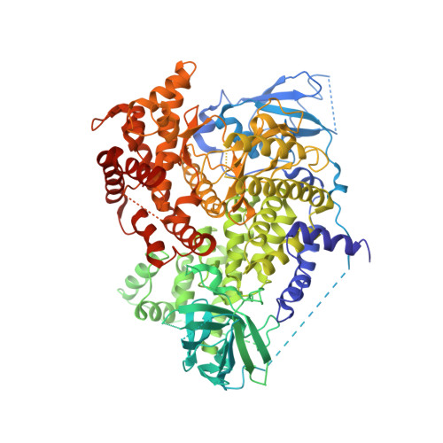
3PRE

Quinazolines with intra-molecular hydrogen bonding scaffold (iMHBS) as PI3K/mTOR dual inhibitors.


Experimental Data Snapshot

  • Method: X-RAY DIFFRACTION
  • Resolution: 2.91 Å
  • R-Value Free: 0.295 
  • R-Value Work: 0.232 
  • R-Value Observed: 0.235 

wwPDB Validation   3D Report Full Report


Ligand Structure Quality Assessment 


This is version 1.3 of the entry. See complete history


Literature

Quinazolines with intra-molecular hydrogen bonding scaffold (iMHBS) as PI3K/mTOR dual inhibitors.

Liu, K.K.Huang, X.Bagrodia, S.Chen, J.H.Greasley, S.Cheng, H.Sun, S.Knighton, D.Rodgers, C.Rafidi, K.Zou, A.Xiao, J.Yan, S.

(2011) Bioorg Med Chem Lett 21: 1270-1274

  • DOI: https://doi.org/10.1016/j.bmcl.2010.12.026
  • Primary Citation of Related Structures:  
    3PRE, 3PRZ, 3PS6

  • PubMed Abstract: 

    Intra-molecular hydrogen bonding was introduced to the quinazoline motif to form a pseudo ring (intra-molecular H-bond scaffold, iMHBS) to mimic our previous published core structures, pyrido[2.3-D]pyrimidin-7-one and pteridinone, as PI3K/mTOR dual inhibitors. This design results in potent PI3K/mTOR dual inhibitors and the purposed intra-molecular hydrogen bonding structure is well supported by co-crystal structure in PI3Kγ enzyme. In addition, a novel synthetic route was developed for these analogs.


  • Organizational Affiliation

    Pfizer Global Research and Development, Chemistry Department, 10770 Science Center Drive, La Jolla, CA 92120, USA. Kevin.K.Liu@pfizer.com


Macromolecules
Find similar proteins by:  (by identity cutoff)  |  3D Structure
Entity ID: 1
MoleculeChains Sequence LengthOrganismDetailsImage
Phosphatidylinositol-4,5-bisphosphate 3-kinase catalytic subunit gamma isoform966Homo sapiensMutation(s): 0 
Gene Names: PIK3CG
EC: 2.7.1.153 (PDB Primary Data), 2.7.1.137 (UniProt), 2.7.1.154 (UniProt), 2.7.11.1 (UniProt)
UniProt & NIH Common Fund Data Resources
Find proteins for P48736 (Homo sapiens)
Explore P48736 
Go to UniProtKB:  P48736
PHAROS:  P48736
GTEx:  ENSG00000105851 
Entity Groups  
Sequence Clusters30% Identity50% Identity70% Identity90% Identity95% Identity100% Identity
UniProt GroupP48736
Sequence Annotations
Expand
  • Reference Sequence
Small Molecules
Ligands 1 Unique
IDChains Name / Formula / InChI Key2D Diagram3D Interactions
3RE
Query on 3RE

Download Ideal Coordinates CCD File 
B [auth A]2-amino-8-(trans-4-methoxycyclohexyl)-4-methyl-6-(1H-pyrazol-3-yl)pyrido[2,3-d]pyrimidin-7(8H)-one
C18 H22 N6 O2
UDXSDTPXGCELRI-HAQNSBGRSA-N
Experimental Data & Validation

Experimental Data

  • Method: X-RAY DIFFRACTION
  • Resolution: 2.91 Å
  • R-Value Free: 0.295 
  • R-Value Work: 0.232 
  • R-Value Observed: 0.235 
  • Space Group: C 1 2 1
Unit Cell:
Length ( Å )Angle ( ˚ )
a = 143.541α = 90
b = 67.276β = 95.77
c = 106.529γ = 90
Software Package:
Software NamePurpose
REFMACrefinement
CNSrefinement
HKL-2000data collection
HKL-2000data reduction
SCALEPACKdata scaling
CNSphasing

Structure Validation

View Full Validation Report


Ligand Structure Quality Assessment 


Entry History 

Deposition Data

Revision History  (Full details and data files)

  • Version 1.0: 2011-02-09
    Type: Initial release
  • Version 1.1: 2011-07-13
    Changes: Version format compliance
  • Version 1.2: 2019-07-17
    Changes: Data collection, Refinement description
  • Version 1.3: 2024-02-21
    Changes: Data collection, Database references, Derived calculations