3Q9U | pdb_00003q9u

In silico and in vitro co-evolution of a high affinity complementary protein-protein interface


Experimental Data Snapshot

  • Method: X-RAY DIFFRACTION
  • Resolution: 2.30 Å
  • R-Value Free: 
    0.318 (Depositor), 0.311 (DCC) 
  • R-Value Work: 
    0.240 (Depositor), 0.236 (DCC) 
  • R-Value Observed: 
    0.244 (Depositor) 

Starting Model: experimental
View more details

wwPDB Validation 3D Report Full Report

Validation slider image for 3Q9U

Ligand Structure Quality Assessment 


This is version 1.3 of the entry. See complete history. 

Literature

A de novo protein binding pair by computational design and directed evolution.

Karanicolas, J., Corn, J.E., Chen, I., Joachimiak, L.A., Dym, O., Peck, S.H., Albeck, S., Unger, T., Hu, W., Liu, G., Delbecq, S., Montelione, G.T., Spiegel, C.P., Liu, D.R., Baker, D.

(2011) Mol Cell 42: 250-260

  • DOI: https://doi.org/10.1016/j.molcel.2011.03.010
  • Primary Citation Related Structures: 
    3Q9N, 3Q9U, 3QA9

  • PubMed Abstract: 

    The de novo design of protein-protein interfaces is a stringent test of our understanding of the principles underlying protein-protein interactions and would enable unique approaches to biological and medical challenges. Here we describe a motif-based method to computationally design protein-protein complexes with native-like interface composition and interaction density. Using this method we designed a pair of proteins, Prb and Pdar, that heterodimerize with a Kd of 130 nM, 1000-fold tighter than any previously designed de novo protein-protein complex. Directed evolution identified two point mutations that improve affinity to 180 pM. Crystal structures of an affinity-matured complex reveal binding is entirely through the designed interface residues. Surprisingly, in the in vitro evolved complex one of the partners is rotated 180° relative to the original design model, yet still maintains the central computationally designed hotspot interaction and preserves the character of many peripheral interactions. This work demonstrates that high-affinity protein interfaces can be created by designing complementary interaction surfaces on two noninteracting partners and underscores remaining challenges.


  • Organizational Affiliation: 
    • Department of Biochemistry, University of Washington, Seattle, WA 98195-7350, USA. johnk@ku.edu

Macromolecule Content 

  • Total Structure Weight: 66.71 kDa 
  • Atom Count: 4,617 
  • Modeled Residue Count: 589 
  • Deposited Residue Count: 598 
  • Unique protein chains: 2

Macromolecules

Find similar proteins by:|  3D Structure
Entity ID: 1
MoleculeChains  Sequence LengthOrganismDetailsImage
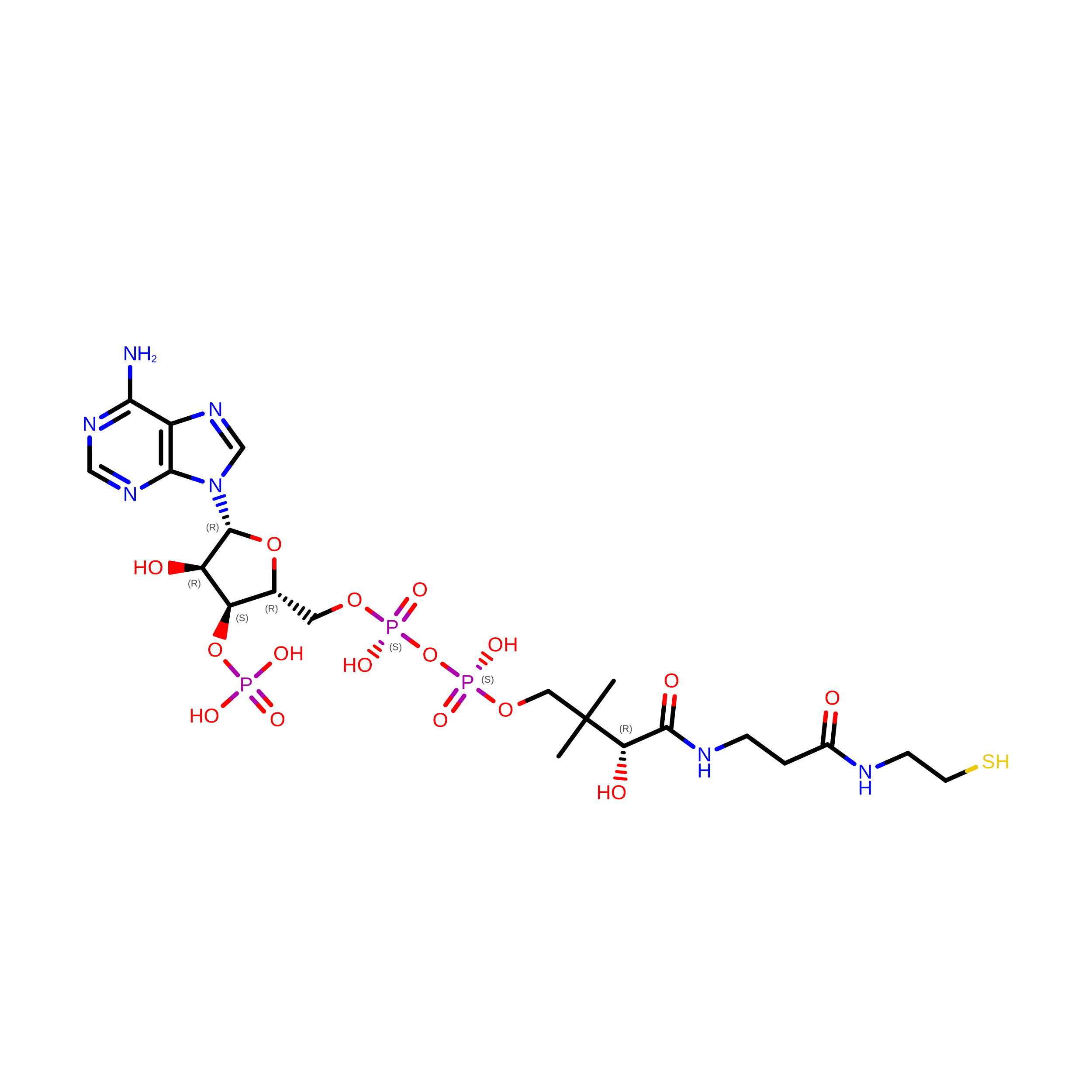
CoA binding protein
A, B
141Escherichia coliMutation(s): 0 
Entity Groups
Sequence Clusters30% Identity50% Identity70% Identity90% Identity95% Identity100% Identity
Sequence Annotations
Expand
Reference Sequence
Find similar proteins by:|  3D Structure
Entity ID: 2
MoleculeChains  Sequence LengthOrganismDetailsImage
consensus ankyrin repeat
C, D
158Escherichia coliMutation(s): 0 
Entity Groups
Sequence Clusters30% Identity50% Identity70% Identity90% Identity95% Identity100% Identity
Sequence Annotations
Expand
Reference Sequence

Experimental Data & Validation

Experimental Data

  • Method: X-RAY DIFFRACTION
  • Resolution: 2.30 Å
  • R-Value Free:  0.318 (Depositor), 0.311 (DCC) 
  • R-Value Work:  0.240 (Depositor), 0.236 (DCC) 
  • R-Value Observed: 0.244 (Depositor) 
Space Group: P 1
Unit Cell:
Length ( Å )Angle ( ˚ )
a = 53.901α = 89.96
b = 57.62β = 90.14
c = 58.23γ = 113.44
Software Package:
Software NamePurpose
PHENIXrefinement

Structure Validation

View Full Validation Report


Ligand Structure Quality Assessment 


Entry History 

Revision History  (Full details and data files)

  • Version 1.0: 2011-04-20
    Type: Initial release
  • Version 1.1: 2011-07-13
    Changes: Version format compliance
  • Version 1.2: 2011-07-20
    Changes: Database references
  • Version 1.3: 2023-09-13
    Changes: Data collection, Database references, Derived calculations, Refinement description