Download Mendeley
A de novo protein binding pair by computational design and directed evolution.
Karanicolas, J., Corn, J.E., Chen, I., Joachimiak, L.A., Dym, O., Peck, S.H., Albeck, S., Unger, T., Hu, W., Liu, G., Delbecq, S., Montelione, G.T., Spiegel, C.P., Liu, D.R., Baker, D.(2011) Mol Cell 42: 250-260
- PubMed: 21458342 Search on PubMedSearch on PubMed Central
- DOI: https://doi.org/10.1016/j.molcel.2011.03.010
- Primary Citation Related Structures:
3Q9N, 3Q9U, 3QA9 - PubMed Abstract:
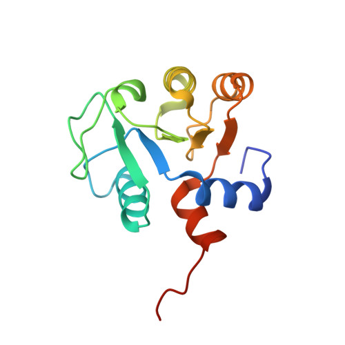
The de novo design of protein-protein interfaces is a stringent test of our understanding of the principles underlying protein-protein interactions and would enable unique approaches to biological and medical challenges. Here we describe a motif-based method to computationally design protein-protein complexes with native-like interface composition and interaction density. Using this method we designed a pair of proteins, Prb and Pdar, that heterodimerize with a Kd of 130 nM, 1000-fold tighter than any previously designed de novo protein-protein complex. Directed evolution identified two point mutations that improve affinity to 180 pM. Crystal structures of an affinity-matured complex reveal binding is entirely through the designed interface residues. Surprisingly, in the in vitro evolved complex one of the partners is rotated 180° relative to the original design model, yet still maintains the central computationally designed hotspot interaction and preserves the character of many peripheral interactions. This work demonstrates that high-affinity protein interfaces can be created by designing complementary interaction surfaces on two noninteracting partners and underscores remaining challenges.
- Department of Biochemistry, University of Washington, Seattle, WA 98195-7350, USA. johnk@ku.edu
Organizational Affiliation: