3QI6 | pdb_00003qi6

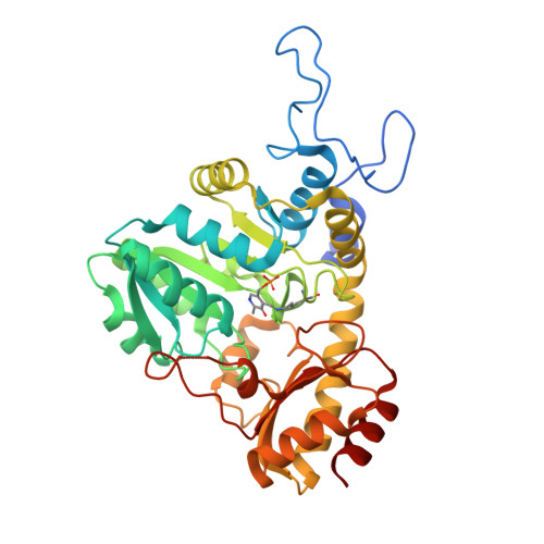
Crystal Structure of Cystathionine gamma-synthase MetB (Cgs) from Mycobacterium ulcerans Agy99


Experimental Data Snapshot

  • Method: X-RAY DIFFRACTION
  • Resolution: 1.91 Å
  • R-Value Free: 
    0.241 (Depositor), 0.206 (DCC) 
  • R-Value Work: 
    0.200 (Depositor) 
  • R-Value Observed: 
    0.202 (Depositor) 

Starting Model: experimental
View more details

wwPDB Validation 3D Report Full Report

Validation slider image for 3QI6

This is version 1.7 of the entry. See complete history. 

Literature

Structure of the cystathionine [gamma]-synthase MetB from Mycobacterium ulcerans

Clifton, M.C., Abendroth, J., Edwards, T.E., Leibly, D.J., Gillespie, A.K., Ferrell, M., Dieterich, S.H., Exley, I., Staker, B.L., Myler, P.J., Van Voorhis, W.C., Stewart, L.J.

(2011) Acta Crystallogr Sect F Struct Biol Cryst Commun 67: 1154-1158

  • DOI: https://doi.org/10.1107/S1744309111029575
  • Primary Citation Related Structures: 
    3QHX, 3QI6

  • PubMed Abstract: 

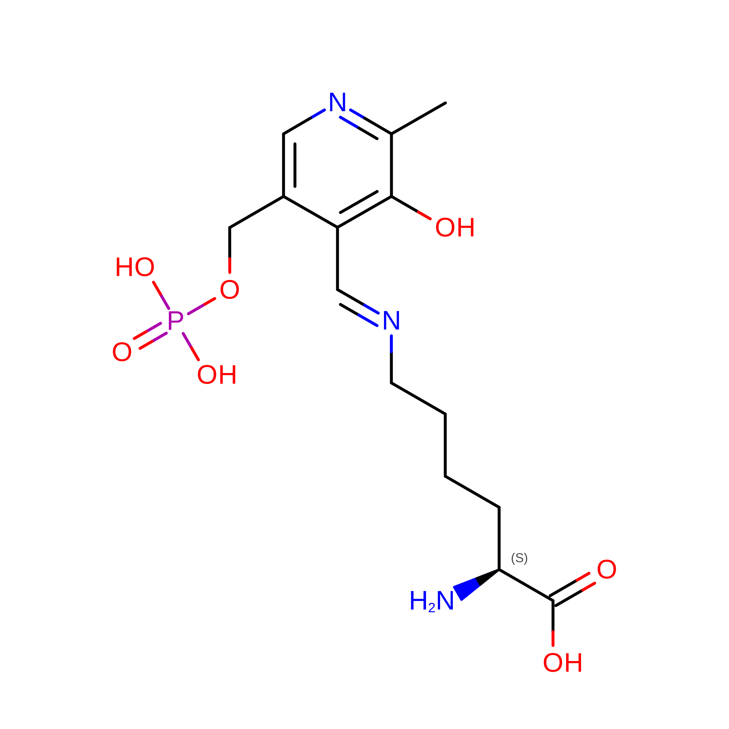
    Cystathionine γ-synthase (CGS) is a transulfurication enzyme that catalyzes the first specific step in L-methionine biosynthesis by the reaction of O(4)-succinyl-L-homoserine and L-cysteine to produce L-cystathionine and succinate. Controlling the first step in L-methionine biosythesis, CGS is an excellent potential drug target. Mycobacterium ulcerans is a slow-growing mycobacterium that is the third most common form of mycobacterial infection, mainly infecting people in Africa, Australia and Southeast Asia. Infected patients display a variety of skin ailments ranging from indolent non-ulcerated lesions as well as ulcerated lesions. Here, the crystal structure of CGS from M. ulcerans covalently linked to the cofactor pyridoxal phosphate (PLP) is reported at 1.9 Å resolution. A second structure contains PLP as well as a highly ordered HEPES molecule in the active site acting as a pseudo-ligand. These results present the first structure of a CGS from a mycobacterium and allow comparison with other CGS enzymes. This is also the first structure reported from the pathogen M. ulcerans.


  • Organizational Affiliation: 
    • Seattle Structural Genomics Center for Infectious Disease (SSGCID), USA. mclifton@embios.com

Macromolecule Content 

  • Total Structure Weight: 165.91 kDa 
  • Atom Count: 12,485 
  • Modeled Residue Count: 1,488 
  • Deposited Residue Count: 1,568 
  • Unique protein chains: 1

Macromolecules

Find similar proteins by:|  3D Structure
Entity ID: 1
MoleculeChains  Sequence LengthOrganismDetailsImage
Cystathionine gamma-synthase MetB (Cgs)
A, B, C, D
392Mycobacterium ulcerans Agy99Mutation(s): 0 
Gene Names: metB, MUL_0201
EC: 2.5.1.48
UniProt
Find proteins for A0PKT3 (Mycobacterium ulcerans (strain Agy99))
Explore A0PKT3 
Go to UniProtKB:  A0PKT3
Entity Groups
Sequence Clusters30% Identity50% Identity70% Identity90% Identity95% Identity100% Identity
UniProt GroupA0PKT3
Sequence Annotations
Expand
Reference Sequence

Small Molecules

Ligands 4 Unique
IDChains Name / Formula / InChI Key2D Diagram3D Interactions
SO4

Query on SO4


Download:Ideal Coordinates CCD File
E [auth A],
F [auth A],
I [auth B],
J [auth B],
L [auth C]
SULFATE ION
O4 S
QAOWNCQODCNURD-UHFFFAOYSA-L
GOL

Query on GOL


Download:Ideal Coordinates CCD File
H [auth A],
K [auth B]
GLYCEROL
C3 H8 O3
PEDCQBHIVMGVHV-UHFFFAOYSA-N
EDO

Query on EDO


Download:Ideal Coordinates CCD File
M [auth D]1,2-ETHANEDIOL
C2 H6 O2
LYCAIKOWRPUZTN-UHFFFAOYSA-N
NA

Query on NA


Download:Ideal Coordinates CCD File
G [auth A]SODIUM ION
Na
FKNQFGJONOIPTF-UHFFFAOYSA-N
Modified Residues  1 Unique
IDChains TypeFormula2D DiagramParent
LLP
Query on LLP
A, B, C, D
L-PEPTIDE LINKINGC14 H22 N3 O7 PLYS

Experimental Data & Validation

Experimental Data

  • Method: X-RAY DIFFRACTION
  • Resolution: 1.91 Å
  • R-Value Free:  0.241 (Depositor), 0.206 (DCC) 
  • R-Value Work:  0.200 (Depositor) 
  • R-Value Observed: 0.202 (Depositor) 
Space Group: P 1 21 1
Unit Cell:
Length ( Å )Angle ( ˚ )
a = 80.956α = 90
b = 106.924β = 113.67
c = 100.307γ = 90
Software Package:
Software NamePurpose
DENZOdata reduction
SCALEPACKdata scaling
PHASERphasing
REFMACrefinement
PDB_EXTRACTdata extraction

Structure Validation

View Full Validation Report


Entry History 

Deposition Data

Revision History  (Full details and data files)

  • Version 1.0: 2011-03-02
    Type: Initial release
  • Version 1.1: 2011-07-13
    Changes: Version format compliance
  • Version 1.2: 2011-09-07
    Changes: Database references, Refinement description
  • Version 1.3: 2012-03-28
    Changes: Database references
  • Version 1.4: 2015-04-22
    Changes: Database references
  • Version 1.5: 2017-11-08
    Changes: Refinement description
  • Version 1.6: 2023-09-13
    Changes: Data collection, Database references, Derived calculations, Refinement description
  • Version 1.7: 2023-12-06
    Changes: Data collection