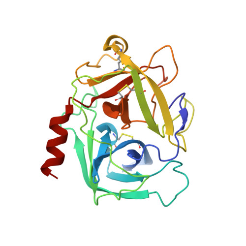
3S0N

Crystal Structure of Human Chymase with Benzimidazolone Inhibitor


Experimental Data Snapshot

  • Method: X-RAY DIFFRACTION
  • Resolution: 1.95 Å
  • R-Value Free: 0.230 
  • R-Value Work: 0.193 

Starting Model: experimental
View more details

wwPDB Validation   3D Report Full Report


Ligand Structure Quality Assessment 


This is version 1.4 of the entry. See complete history


Literature

Benzimidazolone as potent chymase inhibitor: Modulation of reactive metabolite formation in the hydrophobic (P(1)) region.

Lo, H.Y.Nemoto, P.A.Kim, J.M.Hao, M.H.Qian, K.C.Farrow, N.A.Albaugh, D.R.Fowler, D.M.Schneiderman, R.D.Michael August, E.Martin, L.Hill-Drzewi, M.Pullen, S.S.Takahashi, H.De Lombaert, S.

(2011) Bioorg Med Chem Lett 21: 4533-4539

  • DOI: https://doi.org/10.1016/j.bmcl.2011.05.126
  • Primary Citation of Related Structures:  
    3S0N

  • PubMed Abstract: 

    A new class of chymase inhibitor featuring a benzimidazolone core with an acid side chain and a P(1) hydrophobic moiety is described. Incubation of the lead compound with GSH resulted in the formation of a GSH conjugate on the benzothiophene P(1) moiety. Replacement of the benzothiophene with different heterocyclic systems such as indoles and benzoisothiazole is feasible. Among the P(1) replacements, benzoisothiazole prevents the formation of GSH conjugate and an in silico analysis of oxidative potentials agreed with the experimental outcome.


  • Organizational Affiliation

    Boehringer Ingelheim Pharmaceuticals Inc., Medicinal Chemistry, Ridgefield, CT 06877, USA. ho-yin.lo@boehringer-ingelheim.com


Macromolecules
Find similar proteins by:  (by identity cutoff)  |  3D Structure
Entity ID: 1
MoleculeChains Sequence LengthOrganismDetailsImage
Chymase226Homo sapiensMutation(s): 3 
Gene Names: CMA1CYHCYM
EC: 3.4.21.39
UniProt & NIH Common Fund Data Resources
Find proteins for P23946 (Homo sapiens)
Explore P23946 
Go to UniProtKB:  P23946
PHAROS:  P23946
GTEx:  ENSG00000092009 
Entity Groups  
Sequence Clusters30% Identity50% Identity70% Identity90% Identity95% Identity100% Identity
UniProt GroupP23946
Glycosylation
Glycosylation Sites: 2Go to GlyGen: P23946-1
Sequence Annotations
Expand
  • Reference Sequence
Small Molecules
Binding Affinity Annotations 
IDSourceBinding Affinity
0BB PDBBind:  3S0N IC50: 180 (nM) from 1 assay(s)
BindingDB:  3S0N IC50: min: 97, max: 180 (nM) from 2 assay(s)
Experimental Data & Validation

Experimental Data

  • Method: X-RAY DIFFRACTION
  • Resolution: 1.95 Å
  • R-Value Free: 0.230 
  • R-Value Work: 0.193 
  • Space Group: P 43
Unit Cell:
Length ( Å )Angle ( ˚ )
a = 73.83α = 90
b = 73.83β = 90
c = 48.45γ = 90
Software Package:
Software NamePurpose
SCALEPACKdata scaling
CNSrefinement
PDB_EXTRACTdata extraction
CrystalCleardata collection
HKL-2000data reduction
CNXphasing
CNXrefinement

Structure Validation

View Full Validation Report


Ligand Structure Quality Assessment 


Entry History 

Deposition Data

Revision History  (Full details and data files)

  • Version 1.0: 2011-07-20
    Type: Initial release
  • Version 1.1: 2011-08-24
    Changes: Database references
  • Version 1.2: 2020-07-29
    Type: Remediation
    Reason: Carbohydrate remediation
    Changes: Data collection, Database references, Derived calculations, Structure summary
  • Version 1.3: 2023-09-13
    Changes: Data collection, Database references, Refinement description, Structure summary
  • Version 1.4: 2024-10-09
    Changes: Structure summary