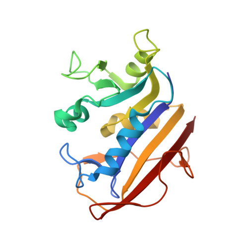
Crystallographic analysis reveals a novel second binding site for trimethoprim in active site double mutants of human dihydrofolate reductase.
Cody, V., Pace, J., Piraino, J., Queener, S.F.(2011) J Struct Biol 176: 52-59
- PubMed: 21684339
- DOI: https://doi.org/10.1016/j.jsb.2011.06.001
- Primary Citation of Related Structures:
3N0H, 3S3V - PubMed Abstract:
In order to produce a more potent replacement for trimethoprim (TMP) used as a therapy for Pneumocystis pneumonia and targets dihydrofolate reductase from Pneumocystis jirovecii (pjDHFR), it is necessary to understand the determinants of potency and selectivity against DHFR from the mammalian host and fungal pathogen cells. To this end, active site residues in human (h) DHFR were replaced with those from pjDHFR. Structural data are reported for two complexes of TMP with the double mutants Gln35Ser/Asn64Phe (Q35S/N64F) and Gln35Lys/Asn64Phe (Q35K/N64F) of hDHFR that unexpectedly show evidence for the binding of two molecules of TMP: one molecule that binds in the normal folate binding site and the second molecule that binds in a novel subpocket site such that the mutated residue Phe64 is involved in van der Waals contacts to the trimethoxyphenyl ring of the second TMP molecule. Kinetic data for the binding of TMP to hDHFR and pjDHFR reveal an 84-fold selectivity of TMP against pjDHFR (K(i) 49 nM) compared to hDHFR (K(i) 4093 nM). Two mutants that contain one substitution from pj--and one from the closely related Pneumocystis carinii DHFR (pcDHFR) (Q35K/N64F and Q35S/N64F) show K(i) values of 593 and 617 nM, respectively; these K(i) values are well above both the K(i) for pjDHFR and are similar to pcDHFR (Q35K/N64F and Q35S/N64F) (305nM). These results suggest that active site residues 35 and 64 play key roles in determining selectivity for pneumocystis DHFR, but that other residues contribute to the unique binding of inhibitors to these enzymes.
- Structural Biology Department, Hauptman Woodward Medical Research Institute, 700 Ellicott St. Buffalo, NY 14203, USA. cody@hwi.buffalo.edu
Organizational Affiliation:


