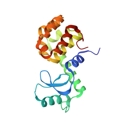
An approach to crystallizing proteins by metal-mediated synthetic symmetrization.
Laganowsky, A., Zhao, M., Soriaga, A.B., Sawaya, M.R., Cascio, D., Yeates, T.O.(2011) Protein Sci 20: 1876-1890
- PubMed: 21898649
- DOI: https://doi.org/10.1002/pro.727
- Primary Citation of Related Structures:
3SB5, 3SB6, 3SB7, 3SB8, 3SB9, 3SBA, 3SBB, 3SER, 3SES, 3SET, 3SEU, 3SEV, 3SEW, 3SEX, 3SEY - PubMed Abstract:
Combining the concepts of synthetic symmetrization with the approach of engineering metal-binding sites, we have developed a new crystallization methodology termed metal-mediated synthetic symmetrization. In this method, pairs of histidine or cysteine mutations are introduced on the surface of target proteins, generating crystal lattice contacts or oligomeric assemblies upon coordination with metal. Metal-mediated synthetic symmetrization greatly expands the packing and oligomeric assembly possibilities of target proteins, thereby increasing the chances of growing diffraction-quality crystals. To demonstrate this method, we designed various T4 lysozyme (T4L) and maltose-binding protein (MBP) mutants and cocrystallized them with one of three metal ions: copper (Cu²⁺, nickel (Ni²⁺), or zinc (Zn²⁺). The approach resulted in 16 new crystal structures--eight for T4L and eight for MBP--displaying a variety of oligomeric assemblies and packing modes, representing in total 13 new and distinct crystal forms for these proteins. We discuss the potential utility of the method for crystallizing target proteins of unknown structure by engineering in pairs of histidine or cysteine residues. As an alternate strategy, we propose that the varied crystallization-prone forms of T4L or MBP engineered in this work could be used as crystallization chaperones, by fusing them genetically to target proteins of interest.
- Institute for Genomics and Proteomics, UCLA-DOE, Los Angeles, California 90095-1570, USA.
Organizational Affiliation:


