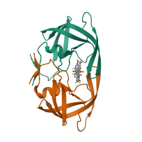
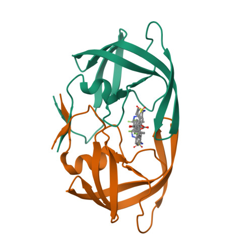
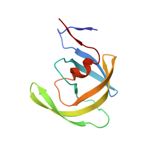
3SPK

Tipranavir in Complex with a Human Immunodeficiency Virus Type 1 Protease Variant


Experimental Data Snapshot

  • Method: X-RAY DIFFRACTION
  • Resolution: 1.24 Å
  • R-Value Free: 0.227 
  • R-Value Work: 0.174 
  • R-Value Observed: 0.177 

wwPDB Validation   3D Report Full Report


Ligand Structure Quality Assessment 


This is version 1.1 of the entry. See complete history


Literature

The higher barrier of darunavir and tipranavir resistance for HIV-1 protease.

Wang, Y.Liu, Z.Brunzelle, J.S.Kovari, I.A.Dewdney, T.G.Reiter, S.J.Kovari, L.C.

(2011) Biochem Biophys Res Commun 412: 737-742

  • DOI: https://doi.org/10.1016/j.bbrc.2011.08.045
  • Primary Citation of Related Structures:  
    3SO9, 3SPK

  • PubMed Abstract: 

    Darunavir and tipranavir are two inhibitors that are active against multi-drug resistant (MDR) HIV-1 protease variants. In this study, the invitro inhibitory efficacy was tested against a MDR HIV-1 protease variant, MDR 769 82T, containing the drug resistance mutations of 46L/54V/82T/84V/90M. Crystallographic and enzymatic studies were performed to examine the mechanism of resistance and the relative maintenance of potency. The key findings are as follows: (i) The MDR protease exhibits decreased susceptibility to all nine HIV-1 protease inhibitors approved by the US Food and Drug Administration (FDA), among which darunavir and tipranavir are the most potent; (ii) the threonine 82 mutation on the protease greatly enhances drug resistance by altering the hydrophobicity of the binding pocket; (iii) darunavir or tipranavir binding facilitates closure of the wide-open flaps of the MDR protease; and (iv) the remaining potency of tipranavir may be preserved by stabilizing the flaps in the inhibitor-protease complex while darunavir maintains its potency by preserving protein main chain hydrogen bonds with the flexible P2 group. These results could provide new insights into drug design strategies to overcome multi-drug resistance of HIV-1 protease variants.


  • Organizational Affiliation

    Department of Biochemistry and Molecular Biology, Wayne State University School of Medicine, Detroit, MI, USA.


Macromolecules
Find similar proteins by:  (by identity cutoff)  |  3D Structure
Entity ID: 1
MoleculeChains Sequence LengthOrganismDetailsImage
HIV-1 protease
A, B
99Human immunodeficiency virus 1Mutation(s): 0 
Gene Names: pol
EC: 3.4.23.16
UniProt
Find proteins for Q000H7 (Human immunodeficiency virus type 1)
Explore Q000H7 
Go to UniProtKB:  Q000H7
Entity Groups  
Sequence Clusters30% Identity50% Identity70% Identity90% Identity95% Identity100% Identity
UniProt GroupQ000H7
Sequence Annotations
Expand
  • Reference Sequence
Small Molecules
Ligands 1 Unique
IDChains Name / Formula / InChI Key2D Diagram3D Interactions
TPV
Query on TPV

Download Ideal Coordinates CCD File 
C [auth A],
D [auth B]
N-(3-{(1R)-1-[(6R)-4-HYDROXY-2-OXO-6-PHENETHYL-6-PROPYL-5,6-DIHYDRO-2H-PYRAN-3-YL]PROPYL}PHENYL)-5-(TRIFLUOROMETHYL)-2-PYRIDINESULFONAMIDE
C31 H33 F3 N2 O5 S
SUJUHGSWHZTSEU-FYBSXPHGSA-N
Binding Affinity Annotations 
IDSourceBinding Affinity
TPV PDBBind:  3SPK IC50: 3.02 (nM) from 1 assay(s)
Experimental Data & Validation

Experimental Data

  • Method: X-RAY DIFFRACTION
  • Resolution: 1.24 Å
  • R-Value Free: 0.227 
  • R-Value Work: 0.174 
  • R-Value Observed: 0.177 
  • Space Group: P 61
Unit Cell:
Length ( Å )Angle ( ˚ )
a = 63.119α = 90
b = 63.119β = 90
c = 83.54γ = 120
Software Package:
Software NamePurpose
HKL-2000data collection
AMoREphasing
REFMACrefinement
DENZOdata reduction
SCALAdata scaling

Structure Validation

View Full Validation Report


Ligand Structure Quality Assessment 


Entry History 

Deposition Data

Revision History  (Full details and data files)

  • Version 1.0: 2011-10-12
    Type: Initial release
  • Version 1.1: 2024-02-28
    Changes: Data collection, Database references, Derived calculations