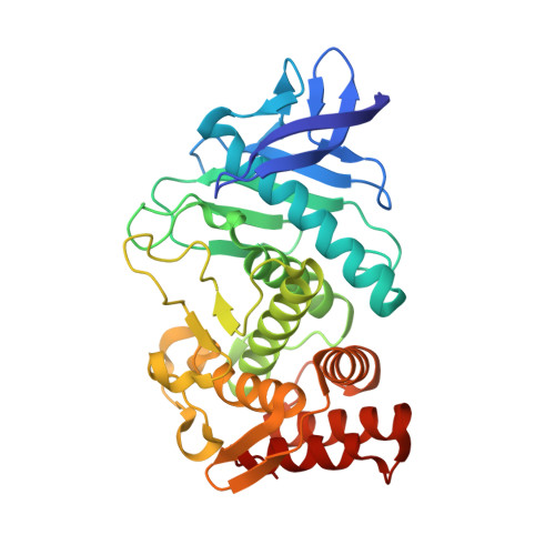
3T73

Thermolysin In Complex With UBTLN22


Experimental Data Snapshot

  • Method: X-RAY DIFFRACTION
  • Resolution: 1.60 Å
  • R-Value Free: 0.166 
  • R-Value Work: 0.143 
  • R-Value Observed: 0.144 

Starting Model: experimental
View more details

wwPDB Validation   3D Report Full Report


Ligand Structure Quality Assessment 


This is version 1.3 of the entry. See complete history


Literature

Water makes the difference: rearrangement of water solvation layer triggers non-additivity of functional group contributions in protein-ligand binding.

Biela, A.Betz, M.Heine, A.Klebe, G.

(2012) ChemMedChem 7: 1423-1434

  • DOI: https://doi.org/10.1002/cmdc.201200206
  • Primary Citation of Related Structures:  
    3T73, 3T74, 3T8F, 3T8G

  • PubMed Abstract: 

    The binding of four congeneric peptide-like thermolysin inhibitors has been studied by high-resolution crystal structure analysis and isothermal titration calorimetry. The ligands differ only by a terminal carboxylate and/or methyl group. A surprising non-additivity of functional group contributions for the carboxylate and/or methyl groups is detected. Adding the methyl first and then the carboxylate group results in a small Gibbs free energy increase and minor enthalpy/entropy partitioning for the first modification, whereas the second involves a strong affinity increase combined with large enthalpy/entropy changes. However, first adding the carboxylate and then the methyl group yields reverse effects: the acidic group attachment now causes minor effects, whereas the added methyl group provokes large changes. As all crystal structures show virtually identical binding modes, affinity changes are related to rearrangements of the first solvation layer next to the S(2)' pocket. About 20-25 water molecules are visible next to the studied complexes. The added COO(-) groups perturb the local water network in both carboxylated complexes, and the attached methyl groups provide favorable interaction sites for water molecules. Apart from one example, a contiguously connected water network between protein and ligand functional groups is observed in all complexes. In the complex with the carboxylated ligand, which still lacks the terminal methyl group, the water network is unfavorably ruptured. This results in a surprising thermodynamic signature showing only a minor affinity increase upon COO(-) group attachment. Because the further added methyl group provides a favorable interaction site for water, the network can be reestablished, and a strong affinity increase with a large enthalpy/entropy signature is then detected.


  • Organizational Affiliation

    Department of Pharmaceutical Chemistry, Philipps University Marburg, Marbacher Weg 6, 35032 Marburg, Germany.


Macromolecules
Find similar proteins by:  (by identity cutoff)  |  3D Structure
Entity ID: 1
MoleculeChains Sequence LengthOrganismDetailsImage
Thermolysin316Bacillus thermoproteolyticusMutation(s): 0 
EC: 3.4.24.27
UniProt
Find proteins for P00800 (Bacillus thermoproteolyticus)
Explore P00800 
Go to UniProtKB:  P00800
Entity Groups  
Sequence Clusters30% Identity50% Identity70% Identity90% Identity95% Identity100% Identity
UniProt GroupP00800
Sequence Annotations
Expand
  • Reference Sequence
Small Molecules
Ligands 5 Unique
IDChains Name / Formula / InChI Key2D Diagram3D Interactions
UBX
Query on UBX

Download Ideal Coordinates CCD File 
B [auth A]N-[(S)-({[(benzyloxy)carbonyl]amino}methyl)(hydroxy)phosphoryl]-N-methyl-L-leucinamide
C16 H26 N3 O5 P
VZBGTFZRCJNJEQ-AWEZNQCLSA-N
GOL
Query on GOL

Download Ideal Coordinates CCD File 
C [auth A],
D [auth A],
E [auth A],
F [auth A]
GLYCEROL
C3 H8 O3
PEDCQBHIVMGVHV-UHFFFAOYSA-N
DMS
Query on DMS

Download Ideal Coordinates CCD File 
G [auth A],
H [auth A],
I [auth A],
J [auth A]
DIMETHYL SULFOXIDE
C2 H6 O S
IAZDPXIOMUYVGZ-UHFFFAOYSA-N
ZN
Query on ZN

Download Ideal Coordinates CCD File 
K [auth A]ZINC ION
Zn
PTFCDOFLOPIGGS-UHFFFAOYSA-N
CA
Query on CA

Download Ideal Coordinates CCD File 
L [auth A],
M [auth A],
N [auth A],
O [auth A]
CALCIUM ION
Ca
BHPQYMZQTOCNFJ-UHFFFAOYSA-N
Experimental Data & Validation

Experimental Data

  • Method: X-RAY DIFFRACTION
  • Resolution: 1.60 Å
  • R-Value Free: 0.166 
  • R-Value Work: 0.143 
  • R-Value Observed: 0.144 
  • Space Group: P 61 2 2
Unit Cell:
Length ( Å )Angle ( ˚ )
a = 92.7α = 90
b = 92.7β = 90
c = 130.1γ = 120
Software Package:
Software NamePurpose
MAR345dtbdata collection
PHASERphasing
PHENIXrefinement
HKL-2000data reduction
HKL-2000data scaling

Structure Validation

View Full Validation Report


Ligand Structure Quality Assessment 


Entry History 

Deposition Data

Revision History  (Full details and data files)

  • Version 1.0: 2012-08-01
    Type: Initial release
  • Version 1.1: 2012-12-12
    Changes: Other
  • Version 1.2: 2014-02-12
    Changes: Database references
  • Version 1.3: 2023-09-13
    Changes: Data collection, Database references, Derived calculations, Refinement description