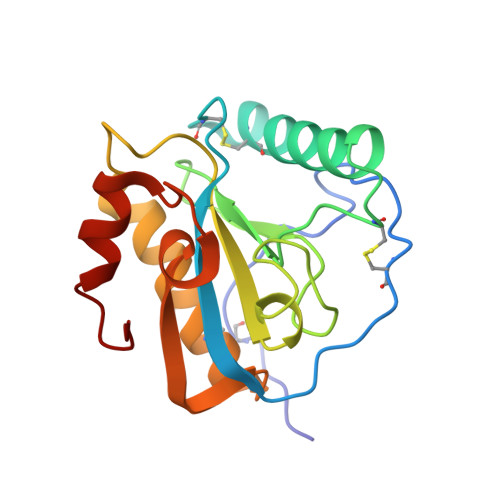
3USX

Crystal structure of PGRP-S complexed with Myristic Acid at 2.28 A resolution


Experimental Data Snapshot

  • Method: X-RAY DIFFRACTION
  • Resolution: 2.28 Å
  • R-Value Free: 0.243 
  • R-Value Work: 0.224 
  • R-Value Observed: 0.224 

Starting Model: experimental
View more details

wwPDB Validation   3D Report Full Report


Ligand Structure Quality Assessment 


This is version 1.3 of the entry. See complete history


Literature

Structural basis of the binding of fatty acids to peptidoglycan recognition protein, PGRP-S through second binding site

Sharma, P.Yamini, S.Dube, D.Singh, A.Mal, G.Pandey, N.Sinha, M.Singh, A.K.Dey, S.Kaur, P.Mitra, D.K.Sharma, S.Singh, T.P.

(2013) Arch Biochem Biophys 529: 1-10

  • DOI: https://doi.org/10.1016/j.abb.2012.11.001
  • Primary Citation of Related Structures:  
    3T2V, 3UIL, 3UMQ, 3USX, 4FNN

  • PubMed Abstract: 

    Short peptidoglycan recognition protein (PGRP-S) is a member of the mammalian innate immune system. PGRP-S from Camelus dromedarius (CPGRP-S) has been shown to bind to lipopolysaccharide (LPS), lipoteichoic acid (LTA) and peptidoglycan (PGN). Its structure consists of four molecules A, B, C and D with ligand binding clefts situated at A-B and C-D contacts. It has been shown that LPS, LTA and PGN bind to CPGRP-S at C-D contact. The cleft at the A-B contact indicated features that suggested a possible binding of fatty acids including mycolic acid of Mycobacterium tuberculosis. Therefore, binding studies of CPGRP-S were carried out with fatty acids, butyric acid, lauric acid, myristic acid, stearic acid and mycolic acid which showed affinities in the range of 10(-5) to 10(-8) M. Structure determinations of the complexes of CPGRP-S with above fatty acids showed that they bound to CPGRP-S in the cleft at the A-B contact. The flow cytometric studies showed that mycolic acid induced the production of pro-inflammatory cytokines, TNF-α and IFN-γ by CD3+ T cells. The concentrations of cytokines increased considerably with increasing concentrations of mycolic acid. However, their levels decreased substantially on adding CPGRP-S.


  • Organizational Affiliation

    Department of Biophysics, All India Institute of Medical Sciences, New Delhi, India.


Macromolecules
Find similar proteins by:  (by identity cutoff)  |  3D Structure
Entity ID: 1
MoleculeChains Sequence LengthOrganismDetailsImage
Peptidoglycan recognition protein 1
A, B, C, D
171Camelus dromedariusMutation(s): 0 
UniProt
Find proteins for Q9GK12 (Camelus dromedarius)
Explore Q9GK12 
Go to UniProtKB:  Q9GK12
Entity Groups  
Sequence Clusters30% Identity50% Identity70% Identity90% Identity95% Identity100% Identity
UniProt GroupQ9GK12
Sequence Annotations
Expand
  • Reference Sequence
Small Molecules
Binding Affinity Annotations 
IDSourceBinding Affinity
MYR PDBBind:  3USX Kd: 9530 (nM) from 1 assay(s)
Experimental Data & Validation

Experimental Data

  • Method: X-RAY DIFFRACTION
  • Resolution: 2.28 Å
  • R-Value Free: 0.243 
  • R-Value Work: 0.224 
  • R-Value Observed: 0.224 
  • Space Group: I 2 2 2
Unit Cell:
Length ( Å )Angle ( ˚ )
a = 89.629α = 90
b = 101.406β = 90
c = 163.069γ = 90
Software Package:
Software NamePurpose
HKL-2000data collection
AMoREphasing
CNSrefinement
DENZOdata reduction
SCALEPACKdata scaling

Structure Validation

View Full Validation Report


Ligand Structure Quality Assessment 


Entry History 

Deposition Data

Revision History  (Full details and data files)

  • Version 1.0: 2012-01-11
    Type: Initial release
  • Version 1.1: 2013-05-15
    Changes: Database references
  • Version 1.2: 2023-11-08
    Changes: Data collection, Database references, Derived calculations, Refinement description
  • Version 1.3: 2024-11-13
    Changes: Structure summary