


3UVR | pdb_00003uvr
3UVR | pdb_00003uvr
Human p38 MAP Kinase in Complex with KM064
- PDB DOI: https://doi.org/10.2210/pdb3UVR/pdb
- Classification: TRANSFERASE/TRANSFERASE INHIBITOR
- Organism(s): Homo sapiens
- Expression System: Escherichia coli
- Mutation(s): No
- Deposited: 2011-11-30 Released: 2012-12-19
Experimental Data Snapshot
- Method: X-RAY DIFFRACTION
- Resolution: 2.10 Å
- R-Value Free: 0.245 (Depositor), 0.243 (DCC)
- R-Value Work: 0.204 (Depositor), 0.204 (DCC)
- R-Value Observed: 0.206 (Depositor)
Starting Model: experimental
View more details

This is version 1.2 of the entry. See complete history.

Macromolecules
Find similar proteins by:
| 3D Structure
Entity ID: 1 | |||||
|---|---|---|---|---|---|
| Molecule | Chains | Sequence Length | Organism | Details | Image |
| Mitogen-activated protein kinase 14 | 360 | Homo sapiens | Mutation(s): 0 Gene Names: MAPK14, CSBP, CSBP1, CSBP2, CSPB1, MXI2 EC: 2.7.11.24 | 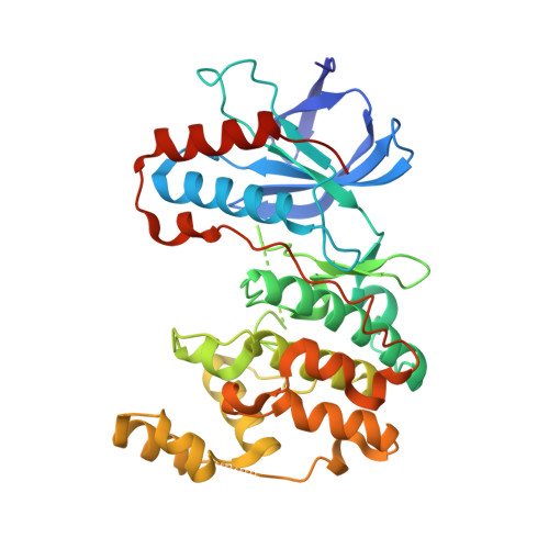 | |
UniProt & NIH Common Fund Data Resources | |||||
PHAROS: Q16539 GTEx: ENSG00000112062 | |||||
Entity Groups | |||||
| Sequence Clusters | 30% Identity50% Identity70% Identity90% Identity95% Identity100% Identity | ||||
| UniProt Group | Q16539 | ||||
Sequence AnnotationsExpand | |||||
Reference Sequence | |||||
Small Molecules
| Ligands 2 Unique | |||||
|---|---|---|---|---|---|
| ID | Chains | Name / Formula / InChI Key | 2D Diagram | 3D Interactions | |
| 06F Download:Ideal Coordinates CCD File | B [auth A] | 1-[3-tert-butyl-1-(4-methylphenyl)-1H-pyrazol-5-yl]-3-{3-[(5-oxo-6,7,8,9-tetrahydro-5H-benzo[7]annulen-2-yl)amino]phenyl}urea C32 H35 N5 O2 ZPGBLWCHXWTWDI-UHFFFAOYSA-N |  | ||
| BOG Download:Ideal Coordinates CCD File | C [auth A], D [auth A] | octyl beta-D-glucopyranoside C14 H28 O6 HEGSGKPQLMEBJL-RKQHYHRCSA-N |  | ||
Experimental Data & Validation
Experimental Data
- Method: X-RAY DIFFRACTION
- Resolution: 2.10 Å
- R-Value Free: 0.245 (Depositor), 0.243 (DCC)
- R-Value Work: 0.204 (Depositor), 0.204 (DCC)
- R-Value Observed: 0.206 (Depositor)
Space Group: P 21 21 21
Unit Cell:
| Length ( Å ) | Angle ( ˚ ) |
|---|---|
| a = 62.858 | α = 90 |
| b = 68.246 | β = 90 |
| c = 73.698 | γ = 90 |
| Software Name | Purpose |
|---|---|
| XSCALE | data scaling |
| PHASER | phasing |
| REFMAC | refinement |
| PDB_EXTRACT | data extraction |
| XDS | data scaling |
Entry History
Deposition Data
- Released Date: 2012-12-19 Deposition Author(s): Richters, A., Mayer-Wrangowski, S.C., Gruetter, C., Rauh, D.
Revision History (Full details and data files)
- Version 1.0: 2012-12-19
Type: Initial release - Version 1.1: 2020-07-29
Type: Remediation
Reason: Carbohydrate remediation
Changes: Data collection, Database references, Derived calculations, Structure summary - Version 1.2: 2023-11-08
Changes: Data collection, Database references, Refinement description, Structure summary


