3V1R

Crystal structures of the reverse transcriptase-associated ribonuclease H domain of XMRV with inhibitor beta-thujaplicinol


Experimental Data Snapshot

  • Method: X-RAY DIFFRACTION
  • Resolution: 2.80 Å
  • R-Value Free: 
    0.277 (Depositor), 0.270 (DCC) 
  • R-Value Work: 
    0.195 (Depositor), 0.190 (DCC) 
  • R-Value Observed: 
    0.205 (Depositor) 

wwPDB Validation   3D Report Full Report


Ligand Structure Quality Assessment 

Created with Raphaël 2.3.0Worse 01 BetterLigand structure goodness of fit to experimental dataBest fitted JTHClick on this verticalbar to view details

This is version 1.2 of the entry. See complete history


Literature

Crystal structures of the reverse transcriptase-associated ribonuclease H domain of xenotropic murine leukemia-virus related virus.

Zhou, D.Chung, S.Miller, M.Le Grice, S.F.Wlodawer, A.

(2012) J Struct Biol 177: 638-645

  • DOI: https://doi.org/10.1016/j.jsb.2012.02.006
  • Primary Citation of Related Structures:  
    3V1O, 3V1Q, 3V1R

  • PubMed Abstract: 

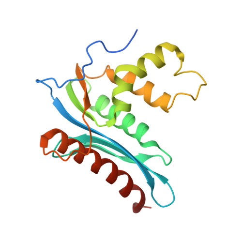
    The ribonuclease H (RNase H) domain of retroviral reverse transcriptase (RT) plays a critical role in the life cycle by degrading the RNA strands of DNA/RNA hybrids. In addition, RNase H activity is required to precisely remove the RNA primers from nascent (-) and (+) strand DNA. We report here three crystal structures of the RNase H domain of xenotropic murine leukemia virus-related virus (XMRV) RT, namely (i) the previously identified construct from which helix C was deleted, (ii) the intact domain, and (iii) the intact domain complexed with an active site α-hydroxytropolone inhibitor. Enzymatic assays showed that the intact RNase H domain retained catalytic activity, whereas the variant lacking helix C was only marginally active, corroborating the importance of this helix for enzymatic activity. Modeling of the enzyme-substrate complex elucidated the essential role of helix C in binding a DNA/RNA hybrid and its likely mode of recognition. The crystal structure of the RNase H domain complexed with β-thujaplicinol clearly showed that coordination by two divalent cations mediates recognition of the inhibitor.


  • Organizational Affiliation

    Protein Structure Section, Macromolecular Crystallography Laboratory, National Cancer Institute at Frederick, Frederick, MD 21702, USA.


Macromolecules
Find similar proteins by:  (by identity cutoff)  |  3D Structure
Entity ID: 1
MoleculeChains Sequence LengthOrganismDetailsImage
Reverse transcriptase/ribonuclease H p80180Xenotropic MuLV-related virus VP35Mutation(s): 0 
Gene Names: gag-polPOL_XMRV3
EC: 3.1.26.4
UniProt
Find proteins for Q2F7J3 (Xenotropic MuLV-related virus (isolate VP35))
Explore Q2F7J3 
Go to UniProtKB:  Q2F7J3
Entity Groups  
Sequence Clusters30% Identity50% Identity70% Identity90% Identity95% Identity100% Identity
UniProt GroupQ2F7J3
Sequence Annotations
Expand
  • Reference Sequence
Small Molecules
Ligands 4 Unique
IDChains Name / Formula / InChI Key2D Diagram3D Interactions
JTH
Query on JTH

Download Ideal Coordinates CCD File 
I [auth A]2,7-dihydroxy-4-(propan-2-yl)cyclohepta-2,4,6-trien-1-one
C10 H12 O3
HVGNSPLLPQWYGC-UHFFFAOYSA-N
MRD
Query on MRD

Download Ideal Coordinates CCD File 
F [auth A](4R)-2-METHYLPENTANE-2,4-DIOL
C6 H14 O2
SVTBMSDMJJWYQN-RXMQYKEDSA-N
SO4
Query on SO4

Download Ideal Coordinates CCD File 
B [auth A],
C [auth A],
D [auth A],
E [auth A]
SULFATE ION
O4 S
QAOWNCQODCNURD-UHFFFAOYSA-L
MN
Query on MN

Download Ideal Coordinates CCD File 
G [auth A],
H [auth A]
MANGANESE (II) ION
Mn
WAEMQWOKJMHJLA-UHFFFAOYSA-N
Experimental Data & Validation

Experimental Data

  • Method: X-RAY DIFFRACTION
  • Resolution: 2.80 Å
  • R-Value Free:  0.277 (Depositor), 0.270 (DCC) 
  • R-Value Work:  0.195 (Depositor), 0.190 (DCC) 
  • R-Value Observed: 0.205 (Depositor) 
Space Group: C 2 2 21
Unit Cell:
Length ( Å )Angle ( ˚ )
a = 53.677α = 90
b = 84.75β = 90
c = 70.817γ = 90
Software Package:
Software NamePurpose
HKL-2000data collection
PHASESphasing
PHENIXrefinement
HKL-2000data reduction
SCALEPACKdata scaling

Structure Validation

View Full Validation Report


Ligand Structure Quality Assessment 

Created with Raphaël 2.3.0Worse 01 BetterLigand structure goodness of fit to experimental dataBest fitted JTHClick on this verticalbar to view details

Entry History 

Deposition Data

Revision History  (Full details and data files)

  • Version 1.0: 2012-03-14
    Type: Initial release
  • Version 1.1: 2012-03-28
    Changes: Database references
  • Version 1.2: 2024-02-28
    Changes: Data collection, Database references, Derived calculations