3WHC

Crystal structure of a transcriptional regulator FadR from Bacillus subtilis in complex with stearoyl-CoA


Experimental Data Snapshot

  • Method: X-RAY DIFFRACTION
  • Resolution: 2.20 Å
  • R-Value Free: 
    0.271 (Depositor), 0.270 (DCC) 
  • R-Value Work: 
    0.239 (Depositor), 0.240 (DCC) 
  • R-Value Observed: 
    0.240 (Depositor) 

Starting Model: experimental
View more details

wwPDB Validation   3D Report Full Report


Ligand Structure Quality Assessment 

Created with Raphaël 2.3.0Worse 01 BetterLigand structure goodness of fit to experimental dataBest fitted ST9Click on this verticalbar to view details

This is version 1.2 of the entry. See complete history


Literature

Structural characterization of a ligand-bound form of Bacillus subtilis FadR involved in the regulation of fatty acid degradation.

Fujihashi, M.Nakatani, T.Hirooka, K.Matsuoka, H.Fujita, Y.Miki, K.

(2014) Proteins 82: 1301-1310

  • DOI: https://doi.org/10.1002/prot.24496
  • Primary Citation of Related Structures:  
    3WHB, 3WHC

  • PubMed Abstract: 

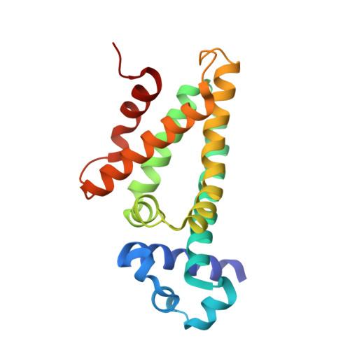
    Bacillus subtilis FadR (FadR(Bs)), a member of the TetR family of bacterial transcriptional regulators, represses five fad operons including 15 genes, most of which are involved in β-oxidation of fatty acids. FadR(Bs) binds to the five FadR(Bs) boxes in the promoter regions and the binding is specifically inhibited by long-chain (C14-C20 ) acyl-CoAs, causing derepression of the fad operons. To elucidate the structural mechanism of this regulator, we have determined the crystal structures of FadR(Bs) proteins prepared with and without stearoyl(C18)-CoA. The crystal structure without adding any ligand molecules unexpectedly includes one small molecule, probably dodecyl(C12)-CoA derived from the Escherichia coli host, in its homodimeric structure. Also, we successfully obtained the structure of the ligand-bound form of the FadR(Bs) dimer by co-crystallization, in which two stearoyl-CoA molecules are accommodated, with the binding mode being essentially equivalent to that of dodecyl-CoA. Although the acyl-chain-binding cavity of FadR(Bs) is mainly hydrophobic, a hydrophilic patch encompasses the C1-C10 carbons of the acyl chain. This accounts for the previous report that the DNA binding of FadR(Bs) is specifically inhibited by the long-chain acyl-CoAs but not by the shorter ones. Structural comparison of the ligand-bound and unliganded subunits of FadR(Bs) revealed three regions around residues 21-31, 61-76, and 106-119 that were substantially changed in response to the ligand binding, and particularly with respect to the movements of Leu108 and Arg109. Site-directed mutagenesis of these residues revealed that Arg109, but not Leu108, is a key residue for maintenance of the DNA-binding affinity of FadR(Bs).


  • Organizational Affiliation

    Department of Chemistry, Graduate School of Science, Kyoto University, Sakyo-ku, Kyoto, 606-8502, Japan.


Macromolecules
Find similar proteins by:  (by identity cutoff)  |  3D Structure
Entity ID: 1
MoleculeChains Sequence LengthOrganismDetailsImage
Fatty acid metabolism regulator protein
A, B, C, D, E
194Bacillus subtilis subsp. subtilis str. 168Mutation(s): 0 
Gene Names: fadRysiABSU28550
UniProt
Find proteins for P94548 (Bacillus subtilis (strain 168))
Explore P94548 
Go to UniProtKB:  P94548
Entity Groups  
Sequence Clusters30% Identity50% Identity70% Identity90% Identity95% Identity100% Identity
UniProt GroupP94548
Sequence Annotations
Expand
  • Reference Sequence
Experimental Data & Validation

Experimental Data

  • Method: X-RAY DIFFRACTION
  • Resolution: 2.20 Å
  • R-Value Free:  0.271 (Depositor), 0.270 (DCC) 
  • R-Value Work:  0.239 (Depositor), 0.240 (DCC) 
  • R-Value Observed: 0.240 (Depositor) 
Space Group: P 21 21 21
Unit Cell:
Length ( Å )Angle ( ˚ )
a = 70.305α = 90
b = 88.762β = 90
c = 199.952γ = 90
Software Package:
Software NamePurpose
REFMACrefinement
PDB_EXTRACTdata extraction
ADSCdata collection
HKL-2000data reduction
HKL-2000data scaling
MOLREPphasing

Structure Validation

View Full Validation Report


Ligand Structure Quality Assessment 

Created with Raphaël 2.3.0Worse 01 BetterLigand structure goodness of fit to experimental dataBest fitted ST9Click on this verticalbar to view details

Entry History 

Deposition Data

Revision History  (Full details and data files)

  • Version 1.0: 2014-03-19
    Type: Initial release
  • Version 1.1: 2017-02-22
    Changes: Database references
  • Version 1.2: 2023-11-08
    Changes: Data collection, Database references, Derived calculations, Refinement description