3ZGC

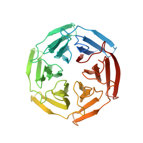
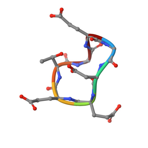
crystal structure of the KEAP1-NEH2 complex


Experimental Data Snapshot

  • Method: X-RAY DIFFRACTION
  • Resolution: 2.20 Å
  • R-Value Free: 0.188 
  • R-Value Work: 0.172 
  • R-Value Observed: 0.173 

Starting Model: experimental
View more details

wwPDB Validation   3D Report Full Report


This is version 1.3 of the entry. See complete history


Literature

Crystal-Contact Engineering to Obtain a Crystal Form of the Kelch Domain of Human Keap1 Suitable for Ligand-Soaking Experiments.

Horer, S.Reinert, D.Ostmann, K.Hoevels, Y.Nar, H.

(2013) Acta Crystallogr Sect F Struct Biol Cryst Commun 69: 592

  • DOI: https://doi.org/10.1107/S174430911301124X
  • Primary Citation of Related Structures:  
    3ZGC, 3ZGD

  • PubMed Abstract: 

    Keap1 is a substrate adaptor protein for a Cul3-dependent ubiquitin ligase complex and plays an important role in the cellular response to oxidative stress. It binds Nrf2 with its Kelch domain and thus triggers the ubiquitinylation and degradation of Nrf2. Oxidative stress prevents the degradation of Nrf2 and leads to the activation of cytoprotective genes. Therefore, Keap1 is an attractive drug target in inflammatory diseases. The support of a medicinal chemistry effort by structural research requires a robust crystallization system in which the crystals are preferably suited for performing soaking experiments. This facilitates the generation of protein-ligand complexes in a routine and high-throughput manner. The structure of human Keap1 has been described previously. In this crystal form, however, the binding site for Nrf2 was blocked by a crystal contact. This interaction was analysed and mutations were introduced to disrupt this crystal contact. One double mutation (E540A/E542A) crystallized in a new crystal form in which the binding site for Nrf2 was not blocked and was accessible to small-molecule ligands. The crystal structures of the apo form of the mutated Keap1 Kelch domain (1.98 Å resolution) and of the complex with an Nrf2-derived peptide obtained by soaking (2.20 Å resolution) are reported.


  • Organizational Affiliation

    Department of Lead Identification and Optimization Support, Boehringer Ingelheim Pharma GmbH and Co KG, Birkendorferstrasse 65, 88400 Biberach, Germany.


Macromolecules
Find similar proteins by:  (by identity cutoff)  |  3D Structure
Entity ID: 1
MoleculeChains Sequence LengthOrganismDetailsImage
KELCH-LIKE ECH-ASSOCIATED PROTEIN 1
A, B
310Homo sapiensMutation(s): 2 
UniProt & NIH Common Fund Data Resources
Find proteins for Q14145 (Homo sapiens)
Explore Q14145 
Go to UniProtKB:  Q14145
PHAROS:  Q14145
GTEx:  ENSG00000079999 
Entity Groups  
Sequence Clusters30% Identity50% Identity70% Identity90% Identity95% Identity100% Identity
UniProt GroupQ14145
Sequence Annotations
Expand
  • Reference Sequence

Find similar proteins by:  Sequence   |   3D Structure  

Entity ID: 2
MoleculeChains Sequence LengthOrganismDetailsImage
NUCLEAR FACTOR ERYTHROID 2-RELATED FACTOR 27Homo sapiensMutation(s): 1 
UniProt & NIH Common Fund Data Resources
Find proteins for Q16236 (Homo sapiens)
Explore Q16236 
Go to UniProtKB:  Q16236
PHAROS:  Q16236
GTEx:  ENSG00000116044 
Entity Groups  
UniProt GroupQ16236
Sequence Annotations
Expand
  • Reference Sequence
Small Molecules
Ligands 1 Unique
IDChains Name / Formula / InChI Key2D Diagram3D Interactions
ACT
Query on ACT

Download Ideal Coordinates CCD File 
D [auth A]
E [auth A]
F [auth A]
G [auth A]
H [auth B]
D [auth A],
E [auth A],
F [auth A],
G [auth A],
H [auth B],
I [auth B],
J [auth B],
K [auth B],
L [auth B],
M [auth B]
ACETATE ION
C2 H3 O2
QTBSBXVTEAMEQO-UHFFFAOYSA-M
Experimental Data & Validation

Experimental Data

  • Method: X-RAY DIFFRACTION
  • Resolution: 2.20 Å
  • R-Value Free: 0.188 
  • R-Value Work: 0.172 
  • R-Value Observed: 0.173 
  • Space Group: P 21 21 21
Unit Cell:
Length ( Å )Angle ( ˚ )
a = 76.103α = 90
b = 76.064β = 90
c = 207.541γ = 90
Software Package:
Software NamePurpose
BUSTERrefinement
XDSdata reduction
SCALAdata scaling
PHASERphasing

Structure Validation

View Full Validation Report


Entry History 

Deposition Data

Revision History  (Full details and data files)

  • Version 1.0: 2013-06-12
    Type: Initial release
  • Version 1.1: 2018-06-20
    Changes: Advisory, Data collection, Derived calculations
  • Version 1.2: 2023-12-20
    Changes: Data collection, Database references, Derived calculations, Other, Refinement description
  • Version 1.3: 2024-10-23
    Changes: Structure summary