4B0A | pdb_00004b0a

The high-resolution structure of yTBP-yTAF1 identifies conserved and competing interaction surfaces in transcriptional activation


Experimental Data Snapshot

  • Method: X-RAY DIFFRACTION
  • Resolution: 1.97 Å
  • R-Value Free: 
    0.223 (Depositor), 0.206 (DCC) 
  • R-Value Work: 
    0.164 (Depositor) 
  • R-Value Observed: 
    0.167 (Depositor) 

Starting Model: experimental
View more details

wwPDB Validation 3D Report Full Report

Validation slider image for 4B0A

This is version 1.4 of the entry. See complete history

Literature

High-Resolution Structure of TBP with Taf1 Reveals Anchoring Patterns in Transcriptional Regulation

Anandapadamanaban, M.Andresen, C.Siponen, M.Kokubo, T.Ikura, M.Moche, M.Sunnerhagen, M.

(2013) Nat Struct Mol Biol 20: 1008

  • DOI: https://doi.org/10.1038/nsmb.2611
  • Primary Citation Related Structures: 
    4B0A

  • PubMed Abstract: 

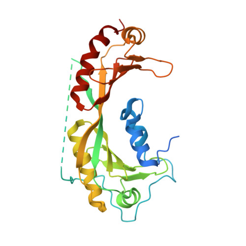
    The general transcription factor TFIID provides a regulatory platform for transcription initiation. Here we present the crystal structure (1.97 Å) and NMR analysis of yeast TAF1 N-terminal domains TAND1 and TAND2 bound to yeast TBP, together with mutational data. We find that yeast TAF1-TAND1, which in itself acts as a transcriptional activator, binds TBP's concave DNA-binding surface by presenting similar anchor residues to TBP as does Mot1 but from a distinct structural scaffold. Furthermore, we show how TAF1-TAND2 uses an aromatic and acidic anchoring pattern to bind a conserved TBP surface groove traversing the basic helix region, and we find highly similar TBP-binding motifs also presented by the structurally distinct TFIIA, Mot1 and Brf1 proteins. Our identification of these anchoring patterns, which can be easily disrupted or enhanced, provides insight into the competitive multiprotein TBP interplay critical to transcriptional regulation.


  • Organizational Affiliation
    • Department of Physics, Chemistry and Biology, Linköping University, Linköping, Sweden.

Macromolecule Content 

  • Total Structure Weight: 31.02 kDa 
  • Atom Count: 2,189 
  • Modeled Residue Count: 237 
  • Deposited Residue Count: 278 
  • Unique protein chains: 1

Macromolecules

Find similar proteins by:|  3D Structure
Entity ID: 1
MoleculeChains  Sequence LengthOrganismDetailsImage
TRANSCRIPTION INITIATION FACTOR TFIID SUBUNIT 1, LINKER, TATA-BOX-BINDING PROTEIN278Saccharomyces cerevisiaeMutation(s): 2 
EC: 2.3.1.48
UniProt
Find proteins for P13393 (Saccharomyces cerevisiae (strain ATCC 204508 / S288c))
Explore P13393 
Go to UniProtKB:  P13393
Find proteins for P46677 (Saccharomyces cerevisiae (strain ATCC 204508 / S288c))
Explore P46677 
Go to UniProtKB:  P46677
Entity Groups
Sequence Clusters30% Identity50% Identity70% Identity90% Identity95% Identity100% Identity
UniProt GroupsP13393P46677
Sequence Annotations
Expand
Reference Sequence

Small Molecules

Ligands 3 Unique
IDChains Name / Formula / InChI Key2D Diagram3D Interactions
GOL

Query on GOL


Download:Ideal Coordinates CCD File
D [auth A]
E [auth A]
F [auth A]
G [auth A]
H [auth A]
D [auth A],
E [auth A],
F [auth A],
G [auth A],
H [auth A],
I [auth A]
GLYCEROL
C3 H8 O3
PEDCQBHIVMGVHV-UHFFFAOYSA-N
CA

Query on CA


Download:Ideal Coordinates CCD File
B [auth A],
C [auth A],
J [auth A]
CALCIUM ION
Ca
BHPQYMZQTOCNFJ-UHFFFAOYSA-N
CL

Query on CL


Download:Ideal Coordinates CCD File
K [auth A]CHLORIDE ION
Cl
VEXZGXHMUGYJMC-UHFFFAOYSA-M

Experimental Data & Validation

Experimental Data

  • Method: X-RAY DIFFRACTION
  • Resolution: 1.97 Å
  • R-Value Free:  0.223 (Depositor), 0.206 (DCC) 
  • R-Value Work:  0.164 (Depositor) 
  • R-Value Observed: 0.167 (Depositor) 
Space Group: P 21 21 21
Unit Cell:
Length ( Å )Angle ( ˚ )
a = 32.894α = 90
b = 74.253β = 90
c = 99.667γ = 90
Software Package:
Software NamePurpose
BUSTERrefinement
XDSdata reduction
SCALAdata scaling
MOLREPphasing

Structure Validation

View Full Validation Report


Entry History 

Deposition Data

Revision History  (Full details and data files)

  • Version 1.0: 2013-07-03
    Type: Initial release
  • Version 1.1: 2013-07-17
    Changes: Database references
  • Version 1.2: 2013-07-24
    Changes: Database references
  • Version 1.3: 2013-08-21
    Changes: Database references
  • Version 1.4: 2023-12-20
    Changes: Data collection, Database references, Derived calculations, Other, Refinement description