4BL3

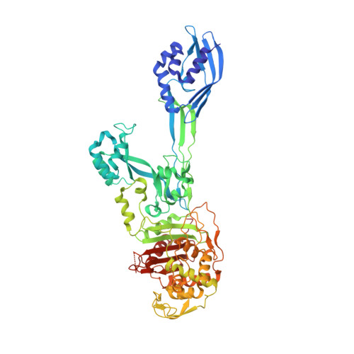
Crystal structure of PBP2a clinical mutant N146K from MRSA


Experimental Data Snapshot

  • Method: X-RAY DIFFRACTION
  • Resolution: 3.00 Å
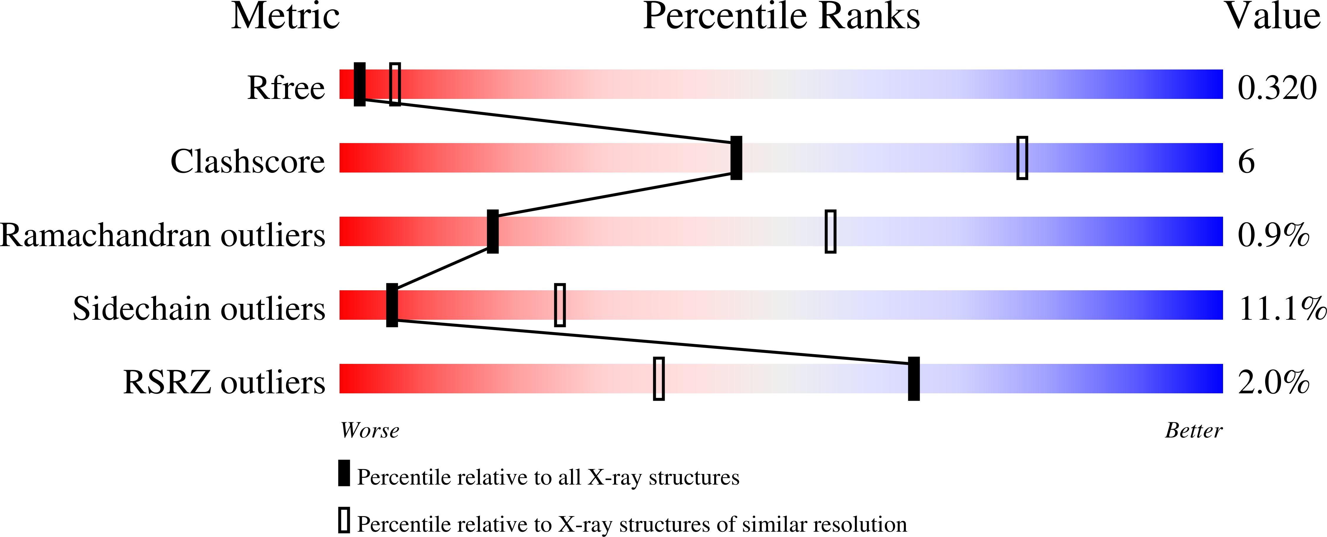
  • R-Value Free: 0.304 
  • R-Value Work: 0.235 
  • R-Value Observed: 0.240 

Starting Model: experimental
View more details

wwPDB Validation   3D Report Full Report


Ligand Structure Quality Assessment 


This is version 1.4 of the entry. See complete history


Literature

Disruption of Allosteric Response as an Unprecedented Mechanism of Resistance to Antibiotics.

Fishovitz, J.Rojas-Altuve, A.Otero, L.H.Dawley, M.Carrasco-Lopez, C.Chang, M.Hermoso, J.A.Mobashery, S.

(2014) J Am Chem Soc 136: 9814

  • DOI: https://doi.org/10.1021/ja5030657
  • Primary Citation of Related Structures:  
    4BL2, 4BL3, 4CPK

  • PubMed Abstract: 

    Ceftaroline, a recently approved β-lactam antibiotic for treatment of infections by methicillin-resistant Staphylococcus aureus (MRSA), is able to inhibit penicillin-binding protein 2a (PBP2a) by triggering an allosteric conformational change that leads to the opening of the active site. The opened active site is now vulnerable to inhibition by a second molecule of ceftaroline, an event that impairs cell-wall biosynthesis and leads to bacterial death. The triggering of the allosteric effect takes place by binding of the first antibiotic molecule 60 Å away from the active site of PBP2a within the core of the allosteric site. We document, by kinetic studies and by determination of three X-ray structures of the mutant variants of PBP2a that result in resistance to ceftaroline, that the effect of these clinical mutants is the disruption of the allosteric trigger in this important protein in MRSA. This is an unprecedented mechanism for antibiotic resistance.


  • Organizational Affiliation

    Department of Chemistry and Biochemistry, University of Notre Dame , Nieuwland Science Hall, Notre Dame, Indiana 46556, United States.


Macromolecules
Find similar proteins by:  (by identity cutoff)  |  3D Structure
Entity ID: 1
MoleculeChains Sequence LengthOrganismDetailsImage
PENICILLIN BINDING PROTEIN 2 PRIME
A, B
640Staphylococcus aureusMutation(s): 1 
EC: 3.4.16.4
UniProt
Find proteins for A0A0H3JPA5 (Staphylococcus aureus (strain Mu50 / ATCC 700699))
Explore A0A0H3JPA5 
Go to UniProtKB:  A0A0H3JPA5
Entity Groups  
Sequence Clusters30% Identity50% Identity70% Identity90% Identity95% Identity100% Identity
UniProt GroupA0A0H3JPA5
Sequence Annotations
Expand
  • Reference Sequence
Small Molecules
Ligands 3 Unique
IDChains Name / Formula / InChI Key2D Diagram3D Interactions
MUR
Query on MUR

Download Ideal Coordinates CCD File 
I [auth A],
O [auth B]
beta-muramic acid
C9 H17 N O7
MSFSPUZXLOGKHJ-KTZFPWNASA-N
CD
Query on CD

Download Ideal Coordinates CCD File 
C [auth A]
D [auth A]
E [auth A]
F [auth A]
J [auth B]
C [auth A],
D [auth A],
E [auth A],
F [auth A],
J [auth B],
K [auth B],
L [auth B]
CADMIUM ION
Cd
WLZRMCYVCSSEQC-UHFFFAOYSA-N
CL
Query on CL

Download Ideal Coordinates CCD File 
G [auth A],
H [auth A],
M [auth B],
N [auth B]
CHLORIDE ION
Cl
VEXZGXHMUGYJMC-UHFFFAOYSA-M
Experimental Data & Validation

Experimental Data

  • Method: X-RAY DIFFRACTION
  • Resolution: 3.00 Å
  • R-Value Free: 0.304 
  • R-Value Work: 0.235 
  • R-Value Observed: 0.240 
  • Space Group: P 21 21 21
Unit Cell:
Length ( Å )Angle ( ˚ )
a = 81.79α = 90
b = 102.39β = 90
c = 187.13γ = 90
Software Package:
Software NamePurpose
BUSTERrefinement
MOSFLMdata reduction
SCALAdata scaling
PHASERphasing

Structure Validation

View Full Validation Report


Ligand Structure Quality Assessment 


Entry History 

Deposition Data

Revision History  (Full details and data files)

  • Version 1.0: 2014-05-21
    Type: Initial release
  • Version 1.1: 2014-09-10
    Changes: Database references
  • Version 1.2: 2017-07-05
    Changes: Data collection
  • Version 1.3: 2020-07-29
    Type: Remediation
    Reason: Carbohydrate remediation
    Changes: Data collection, Derived calculations, Other, Structure summary
  • Version 1.4: 2023-12-20
    Changes: Data collection, Database references, Refinement description, Structure summary