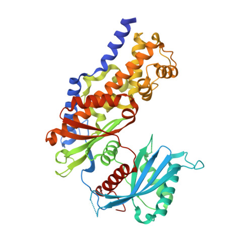
4DCH

Insights into Glucokinase Activation Mechanism: Observation of Multiple Distinct Protein Conformations


Experimental Data Snapshot

  • Method: X-RAY DIFFRACTION
  • Resolution: 1.79 Å
  • R-Value Free: 0.231 
  • R-Value Work: 0.198 
  • R-Value Observed: 0.200 

wwPDB Validation   3D Report Full Report


Ligand Structure Quality Assessment 


This is version 1.5 of the entry. See complete history


Literature

Insights into Mechanism of Glucokinase Activation: OBSERVATION OF MULTIPLE DISTINCT PROTEIN CONFORMATIONS.

Liu, S.Ammirati, M.J.Song, X.Knafels, J.D.Zhang, J.Greasley, S.E.Pfefferkorn, J.A.Qiu, X.

(2012) J Biol Chem 287: 13598-13610

  • DOI: https://doi.org/10.1074/jbc.M111.274126
  • Primary Citation of Related Structures:  
    3VEV, 3VEY, 3VF6, 4DCH, 4DHY

  • PubMed Abstract: 

    Human glucokinase (GK) is a principal regulating sensor of plasma glucose levels. Mutations that inactivate GK are linked to diabetes, and mutations that activate it are associated with hypoglycemia. Unique kinetic properties equip GK for its regulatory role: although it has weak basal affinity for glucose, positive cooperativity in its binding of glucose causes a rapid increase in catalytic activity when plasma glucose concentrations rise above euglycemic levels. In clinical trials, small molecule GK activators (GKAs) have been efficacious in lowering plasma glucose and enhancing glucose-stimulated insulin secretion, but they carry a risk of overly activating GK and causing hypoglycemia. The theoretical models proposed to date attribute the positive cooperativity of GK to the existence of distinct protein conformations that interconvert slowly and exhibit different affinities for glucose. Here we report the respective crystal structures of the catalytic complex of GK and of a GK-glucose complex in a wide open conformation. To assess conformations of GK in solution, we also carried out small angle x-ray scattering experiments. The results showed that glucose dose-dependently converts GK from an apo conformation to an active open conformation. Compared with wild type GK, activating mutants required notably lower concentrations of glucose to be converted to the active open conformation. GKAs decreased the level of glucose required for GK activation, and different compounds demonstrated distinct activation profiles. These results lead us to propose a modified mnemonic model to explain cooperativity in GK. Our findings may offer new approaches for designing GKAs with reduced hypoglycemic risk.


  • Organizational Affiliation

    Structural Biology and Biophysics, Pfizer Groton Laboratories, Groton, Connecticut 06340, USA. shenping.liu@pfizer.com


Macromolecules
Find similar proteins by:  (by identity cutoff)  |  3D Structure
Entity ID: 1
MoleculeChains Sequence LengthOrganismDetailsImage
Glucokinase473Homo sapiensMutation(s): 0 
Gene Names: GCK
EC: 2.7.1.2 (PDB Primary Data), 2.7.1.1 (UniProt)
UniProt & NIH Common Fund Data Resources
Find proteins for P35557 (Homo sapiens)
Explore P35557 
Go to UniProtKB:  P35557
PHAROS:  P35557
GTEx:  ENSG00000106633 
Entity Groups  
Sequence Clusters30% Identity50% Identity70% Identity90% Identity95% Identity100% Identity
UniProt GroupP35557
Sequence Annotations
Expand
  • Reference Sequence
Small Molecules
Binding Affinity Annotations 
IDSourceBinding Affinity
4DC BindingDB:  4DCH Kd: 500 (nM) from 1 assay(s)
EC50: min: 500, max: 2.67e+4 (nM) from 5 assay(s)
Experimental Data & Validation

Experimental Data

  • Method: X-RAY DIFFRACTION
  • Resolution: 1.79 Å
  • R-Value Free: 0.231 
  • R-Value Work: 0.198 
  • R-Value Observed: 0.200 
  • Space Group: P 1 21 1
Unit Cell:
Length ( Å )Angle ( ˚ )
a = 49.917α = 90
b = 85.794β = 104.4
c = 72.939γ = 90
Software Package:
Software NamePurpose
SCALEPACKdata scaling
MOLREPphasing
REFMACrefinement
PDB_EXTRACTdata extraction
Web-Icedata collection
DENZOdata reduction

Structure Validation

View Full Validation Report


Ligand Structure Quality Assessment 


Entry History 

Deposition Data

Revision History  (Full details and data files)

  • Version 1.0: 2012-02-08
    Type: Initial release
  • Version 1.1: 2012-04-11
    Changes: Database references
  • Version 1.2: 2012-05-09
    Changes: Database references
  • Version 1.3: 2017-11-15
    Changes: Refinement description
  • Version 1.4: 2020-07-29
    Type: Remediation
    Reason: Carbohydrate remediation
    Changes: Data collection, Database references, Derived calculations, Structure summary
  • Version 1.5: 2024-02-28
    Changes: Data collection, Database references, Structure summary