4DDP | pdb_00004ddp

crystal structure of Beclin 1 evolutionarily conserved domain(ECD)


Experimental Data Snapshot

  • Method: X-RAY DIFFRACTION
  • Resolution: 1.55 Å
  • R-Value Free: 
    0.203 (Depositor), 0.205 (DCC) 
  • R-Value Work: 
    0.175 (Depositor), 0.184 (DCC) 
  • R-Value Observed: 
    0.177 (Depositor) 

wwPDB Validation 3D Report Full Report

Validation slider image for 4DDP

This is version 1.1 of the entry. See complete history

Literature

Crystal structure and biochemical analyses reveal Beclin 1 as a novel membrane binding protein

Huang, W.Choi, W.Hu, W.Mi, N.Guo, Q.Ma, M.Liu, M.Tian, Y.Lu, P.Wang, F.L.Deng, H.Liu, L.Gao, N.Yu, L.Shi, Y.

(2012) Cell Res 

  • DOI: https://doi.org/10.1038/cr.2012.24
  • Primary Citation Related Structures: 
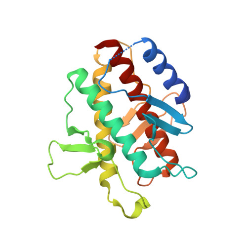
    4DDP

  • PubMed Abstract: 

    The Beclin 1 gene is a haplo-insufficient tumor suppressor and plays an essential role in autophagy. However, the molecular mechanism by which Beclin 1 functions remains largely unknown. Here we report the crystal structure of the evolutionarily conserved domain (ECD) of Beclin 1 at 1.6 Å resolution. Beclin 1 ECD exhibits a previously unreported fold, with three structural repeats arranged symmetrically around a central axis. Beclin 1 ECD defines a novel class of membrane-binding domain, with a strong preference for lipid membrane enriched with cardiolipin. The tip of a surface loop in Beclin 1 ECD, comprising three aromatic amino acids, acts as a hydrophobic finger to associate with lipid membrane, consequently resulting in the deformation of membrane and liposomes. Mutation of these aromatic residues rendered Beclin 1 unable to stably associate with lipid membrane in vitro and unable to fully rescue autophagy in Beclin 1-knockdown cells in vivo. These observations form an important framework for deciphering the biological functions of Beclin 1.


  • Organizational Affiliation
    • Tsinghua-Peking Joint Center for Life Sciences, Tsinghua University, Beijing 100084, China.

Macromolecule Content 

  • Total Structure Weight: 24.46 kDa 
  • Atom Count: 1,705 
  • Modeled Residue Count: 195 
  • Deposited Residue Count: 210 
  • Unique protein chains: 1

Macromolecules

Find similar proteins by:|  3D Structure
Entity ID: 1
MoleculeChains  Sequence LengthOrganismDetailsImage
Beclin-1210Homo sapiensMutation(s): 0 
Gene Names: BECN1GT197
UniProt & NIH Common Fund Data Resources
Find proteins for Q14457 (Homo sapiens)
Explore Q14457 
Go to UniProtKB:  Q14457
PHAROS:  Q14457
GTEx:  ENSG00000126581 
Entity Groups
Sequence Clusters30% Identity50% Identity70% Identity90% Identity95% Identity100% Identity
UniProt GroupQ14457
Sequence Annotations
Expand
Reference Sequence

Experimental Data & Validation

Experimental Data

  • Method: X-RAY DIFFRACTION
  • Resolution: 1.55 Å
  • R-Value Free:  0.203 (Depositor), 0.205 (DCC) 
  • R-Value Work:  0.175 (Depositor), 0.184 (DCC) 
  • R-Value Observed: 0.177 (Depositor) 
Space Group: C 1 2 1
Unit Cell:
Length ( Å )Angle ( ˚ )
a = 80.746α = 90
b = 33.563β = 113.49
c = 74.765γ = 90
Software Package:
Software NamePurpose
CBASSdata collection
PHASERphasing
PHENIXrefinement
HKL-2000data reduction
HKL-2000data scaling

Structure Validation

View Full Validation Report


Entry History 

Deposition Data

Revision History  (Full details and data files)

  • Version 1.0: 2012-02-22
    Type: Initial release
  • Version 1.1: 2024-03-20
    Changes: Data collection, Database references