4FWW | pdb_00004fww

Crystal structure of the Sema-PSI extracellular domains of human RON receptor tyrosine kinase


Experimental Data Snapshot

  • Method: X-RAY DIFFRACTION
  • Resolution: 1.85 Å
  • R-Value Free: 
    0.232 (Depositor), 0.238 (DCC) 
  • R-Value Work: 
    0.191 (Depositor), 0.208 (DCC) 
  • R-Value Observed: 
    0.193 (Depositor) 

Starting Model: experimental
View more details

wwPDB Validation 3D Report Full Report

Validation slider image for 4FWW

This is version 2.2 of the entry. See complete history

Literature

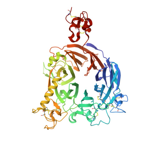
Crystal Structure of the Sema-PSI Extracellular Domain of Human RON Receptor Tyrosine Kinase.

Chao, K.L.Tsai, I.W.Chen, C.Herzberg, O.

(2012) PLoS One 7: e41912-e41912

  • DOI: https://doi.org/10.1371/journal.pone.0041912
  • Primary Citation Related Structures: 
    4FWW

  • PubMed Abstract: 

    Human RON (Recepteur d'Origine Nantais) receptor tyrosine kinase is a cell surface receptor for Macrophage Stimulating Protein (MSP). RON mediates signal transduction pathways that regulate cell adhesion, invasion, motility and apoptosis processes. Elevated levels of RON and its alternatively spliced variants are implicated in the progression and metastasis of tumor cells. The binding of MSP α/β heterodimer to the extracellular region of RON receptor induces receptor dimerization and activation by autophosphorylation of the intracellular kinase domains. The ectodomain of RON, containing the ligand recognition and dimerization domains, is composed of a semaphorin (Sema), Plexins-Semaphorins-Integrins domain (PSI), and four Immunoglobulins-Plexins-Transcription factor (IPT) domains. High affinity association between MSP and RON is mediated by the interaction between MSP β-chain and RON Sema, although RON activation requires intact RON and MSP proteins. Here, we report the structure of RON Sema-PSI domains at 1.85 Å resolution. RON Sema domain adopts a seven-bladed β-propeller fold, followed by disulfide bond rich, cysteine-knot PSI motif. Comparison with the homologous Met receptor tyrosine kinase reveals that RON Sema-PSI contains distinguishing secondary structural features. These define the receptors' exclusive selectivity towards their respective ligands, RON for MSP and Met for HGF. The RON Sema-PSI crystal packing generates a homodimer with interface formed by the Sema domain. Mapping of the dimer interface using the RON homology to Met, MSP homology to Hepatocyte Growth Factor (HGF), and the structure of the Met/HGF complex shows the dimer interface overlapping with the putative MSPβ binding site. The crystallographically determined RON Sema-PSI homodimer may represent the dimer assembly that occurs during ligand-independent receptor activation and/or the inhibition of the constitutive activity of RONΔ160 splice variant by the soluble RON splice variant, RONΔ85.


  • Organizational Affiliation
    • Institute for Bioscience and Biotechnology Research, University of Maryland, Rockville, Maryland, United States of America.

Macromolecule Content 

  • Total Structure Weight: 58.09 kDa 
  • Atom Count: 4,330 
  • Modeled Residue Count: 513 
  • Deposited Residue Count: 527 
  • Unique protein chains: 1

Macromolecules

Find similar proteins by:|  3D Structure
Entity ID: 1
MoleculeChains  Sequence LengthOrganismDetailsImage
Macrophage-stimulating protein receptor527Homo sapiensMutation(s): 0 
Gene Names: MST1RPTK8RON
EC: 2.7.10.1
UniProt & NIH Common Fund Data Resources
Find proteins for Q04912 (Homo sapiens)
Explore Q04912 
Go to UniProtKB:  Q04912
PHAROS:  Q04912
GTEx:  ENSG00000164078 
Entity Groups
Sequence Clusters30% Identity50% Identity70% Identity90% Identity95% Identity100% Identity
UniProt GroupQ04912
Glycosylation
Glycosylation Sites: 1Go to GlyGen: Q04912-1
Sequence Annotations
Expand
Reference Sequence

Oligosaccharides

Help  
Entity ID: 2
MoleculeChains Length2D Diagram GlycosylationD Interactions
beta-D-mannopyranose-(1-3)-beta-D-mannopyranose-(1-4)-2-acetamido-2-deoxy-beta-D-glucopyranose-(1-4)-2-acetamido-2-deoxy-beta-D-glucopyranose
B
4N-Glycosylation
Glycosylation Resources
GlyTouCan: G31886NL
GlyCosmos: G31886NL
GlyGen: G31886NL

Small Molecules

Ligands 5 Unique
IDChains Name / Formula / InChI Key2D Diagram3D Interactions
PEG

Query on PEG


Download:Ideal Coordinates CCD File
R [auth A]DI(HYDROXYETHYL)ETHER
C4 H10 O3
MTHSVFCYNBDYFN-UHFFFAOYSA-N
SO4

Query on SO4


Download:Ideal Coordinates CCD File
C [auth A],
D [auth A]
SULFATE ION
O4 S
QAOWNCQODCNURD-UHFFFAOYSA-L
GOL

Query on GOL


Download:Ideal Coordinates CCD File
J [auth A],
K [auth A],
L [auth A],
M [auth A]
GLYCEROL
C3 H8 O3
PEDCQBHIVMGVHV-UHFFFAOYSA-N
EDO

Query on EDO


Download:Ideal Coordinates CCD File
N [auth A],
O [auth A],
P [auth A],
Q [auth A]
1,2-ETHANEDIOL
C2 H6 O2
LYCAIKOWRPUZTN-UHFFFAOYSA-N
ACT

Query on ACT


Download:Ideal Coordinates CCD File
E [auth A],
F [auth A],
G [auth A],
H [auth A],
I [auth A]
ACETATE ION
C2 H3 O2
QTBSBXVTEAMEQO-UHFFFAOYSA-M

Experimental Data & Validation

Experimental Data

  • Method: X-RAY DIFFRACTION
  • Resolution: 1.85 Å
  • R-Value Free:  0.232 (Depositor), 0.238 (DCC) 
  • R-Value Work:  0.191 (Depositor), 0.208 (DCC) 
  • R-Value Observed: 0.193 (Depositor) 
Space Group: C 1 2 1
Unit Cell:
Length ( Å )Angle ( ˚ )
a = 106.4α = 90
b = 73.3β = 97.4
c = 87.9γ = 90
Software Package:
Software NamePurpose
JBluIce-EPICSdata collection
PHASERphasing
REFMACrefinement
XDSdata reduction
XSCALEdata scaling

Structure Validation

View Full Validation Report


Entry History 

Deposition Data

Revision History  (Full details and data files)

  • Version 1.0: 2012-08-08
    Type: Initial release
  • Version 1.1: 2012-08-22
    Changes: Database references
  • Version 1.2: 2017-11-15
    Changes: Advisory, Refinement description
  • Version 2.0: 2020-07-29
    Type: Remediation
    Reason: Carbohydrate remediation
    Changes: Atomic model, Data collection, Database references, Derived calculations, Structure summary
  • Version 2.1: 2023-09-13
    Changes: Advisory, Data collection, Database references, Refinement description, Structure summary
  • Version 2.2: 2024-11-06
    Changes: Structure summary