4H4L | pdb_00004h4l

Crystal Structure of ternary complex of HutP(HutP-L-His-Zn)


Experimental Data Snapshot

  • Method: X-RAY DIFFRACTION
  • Resolution: 2.50 Å
  • R-Value Free: 
    0.298 (Depositor), 0.297 (DCC) 
  • R-Value Work: 
    0.219 (Depositor), 0.220 (DCC) 
  • R-Value Observed: 
    0.223 (Depositor) 

Starting Model: experimental
View more details

wwPDB Validation 3D Report Full Report

Validation slider image for 4H4L

Ligand Structure Quality Assessment 


This is version 1.3 of the entry. See complete history

Literature

Alternative binding modes of l-histidine guided by metal ions for the activation of the antiterminator protein HutP of Bacillus subtilis.

Dhakshnamoorthy, B.Mizuno, H.Kumar, P.K.R.

(2013) J Struct Biol 183: 512-518

  • DOI: https://doi.org/10.1016/j.jsb.2013.05.019
  • Primary Citation Related Structures: 
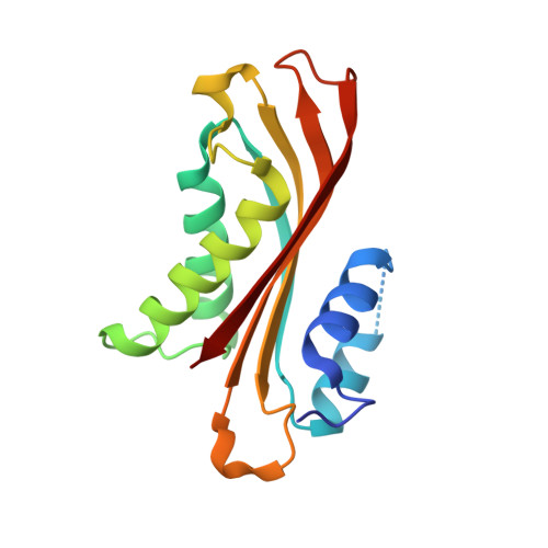
    4H4L

  • PubMed Abstract: 

    Anti-terminator proteins control gene expression by recognizing control signals within cognate transcripts and then preventing transcription termination. HutP is such a regulatory protein that regulates the expression of the histidine utilization (hut) operon in Bacillus subtilis by binding to cis-acting regulatory sequences in hut mRNAs. During the anti-termination process, l-histidine and a divalent ion are required for hutP to bind to the specific sequence within the hut mRNA. Our previous crystal structure of the HutP-l-histidine-Mg(2+)-RNA ternary complex demonstrated that the l-histidine ligand and Mg(2+) bind together such that the backbone nitrogen and carboxyl oxygen of l-histidine coordinate with Mg(2+). In addition to the Mg(2+), other divalent ions are also known to efficiently support the l-histidine-dependent anti-termination of the hut operon, and the best divalent ion is Zn(2+). In this study, we determined the crystal structure of the HutP-l-histidine-Zn(2+) complex and found that the orientation of l-histidine coordinated to Zn(2+) is reversed relative to that of l-histidine coordinated to Mg(2+), i.e., the imidazole side chain nitrogen of l-histidine coordinates to Zn(2+). This alternative binding mode of the l-histidine ligand to a divalent ion provides further insight into the mechanisms responsible for the activation of RNA binding during the hut anti-termination process.


  • Organizational Affiliation
    • RNA Processing Group, Biomedical Research Institute, National Institute of Advanced Industrial Science and Technology, Central-6, 1-1-1 Higashi, Tsukuba 305-8566, Japan.

Macromolecule Content 

  • Total Structure Weight: 197.45 kDa 
  • Atom Count: 13,047 
  • Modeled Residue Count: 1,706 
  • Deposited Residue Count: 1,776 
  • Unique protein chains: 1

Macromolecules

Find similar proteins by:|  3D Structure
Entity ID: 1
MoleculeChains  Sequence LengthOrganismDetailsImage
Hut operon positive regulatory protein
A, B, C, D, E
A, B, C, D, E, F, G, H, I, J, K, L
148Bacillus subtilis subsp. subtilis str. 168Mutation(s): 1 
Gene Names: BSU39340hutP
UniProt
Find proteins for P10943 (Bacillus subtilis (strain 168))
Explore P10943 
Go to UniProtKB:  P10943
Entity Groups
Sequence Clusters30% Identity50% Identity70% Identity90% Identity95% Identity100% Identity
UniProt GroupP10943
Sequence Annotations
Expand
Reference Sequence

Small Molecules

Ligands 2 Unique
IDChains Name / Formula / InChI Key2D Diagram3D Interactions
HIS

Query on HIS


Download:Ideal Coordinates CCD File
AA [auth G]
CA [auth I]
EA [auth J]
GA [auth J]
IA [auth L]
AA [auth G],
CA [auth I],
EA [auth J],
GA [auth J],
IA [auth L],
M [auth A],
O [auth B],
Q [auth C],
S [auth D],
U [auth D],
W [auth F],
Y [auth G]
HISTIDINE
C6 H10 N3 O2
HNDVDQJCIGZPNO-YFKPBYRVSA-O
ZN

Query on ZN


Download:Ideal Coordinates CCD File
BA [auth H]
DA [auth I]
FA [auth J]
HA [auth K]
JA [auth L]
BA [auth H],
DA [auth I],
FA [auth J],
HA [auth K],
JA [auth L],
N [auth A],
P [auth B],
R [auth C],
T [auth D],
V [auth E],
X [auth F],
Z [auth G]
ZINC ION
Zn
PTFCDOFLOPIGGS-UHFFFAOYSA-N

Experimental Data & Validation

Experimental Data

  • Method: X-RAY DIFFRACTION
  • Resolution: 2.50 Å
  • R-Value Free:  0.298 (Depositor), 0.297 (DCC) 
  • R-Value Work:  0.219 (Depositor), 0.220 (DCC) 
  • R-Value Observed: 0.223 (Depositor) 
Space Group: P 3
Unit Cell:
Length ( Å )Angle ( ˚ )
a = 129.53α = 90
b = 129.53β = 90
c = 76.67γ = 120
Software Package:
Software NamePurpose
ADSCdata collection
PHASERphasing
REFMACrefinement
HKL-2000data reduction
HKL-2000data scaling

Structure Validation

View Full Validation Report


Ligand Structure Quality Assessment 


Entry History 

Deposition Data

Revision History  (Full details and data files)

  • Version 1.0: 2013-09-04
    Type: Initial release
  • Version 1.1: 2014-09-10
    Changes: Database references
  • Version 1.2: 2017-11-15
    Changes: Advisory, Refinement description
  • Version 1.3: 2023-11-08
    Changes: Advisory, Data collection, Database references, Derived calculations, Refinement description