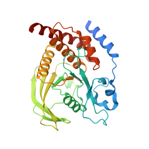
4HJQ

SHP-1 catalytic domain WPD loop closed


Experimental Data Snapshot

  • Method: X-RAY DIFFRACTION
  • Resolution: 1.80 Å
  • R-Value Free: 0.202 
  • R-Value Work: 0.165 
  • R-Value Observed: 0.167 

Starting Model: experimental
View more details

wwPDB Validation   3D Report Full Report


This is version 1.3 of the entry. See complete history


Literature

SHP Family Protein Tyrosine Phosphatases Adopt Canonical Active-Site Conformations in the Apo and Phosphate-Bound States.

Alicea-Velazquez, N.L.Boggon, T.J.

(2013) Protein Pept Lett 20: 1039-1048

  • DOI: https://doi.org/10.2174/09298665113209990041
  • Primary Citation of Related Structures:  
    4HJP, 4HJQ

  • PubMed Abstract: 

    Protein tyrosine phosphatase (PTP) catalytic domains undergo a series of conformational changes in order to mediate dephosphorylation of their tyrosine phosphorylated substrates. An important conformational change occurs in the Tryptophan-Proline-Aspartic acid (WPD) loop, which contains the conserved catalytic aspartate. Upon substrate binding, the WPD loop transitions from the 'open' to the 'closed' state, thus allowing optimal positioning of the catalytic aspartate for substrate dephosphorylation. The dynamics of WPD loop conformational changes have previously been studied for PTP1B, HePTP, and the bacterial phosphatase YopH, but have not yet been comprehensively studied for the nonreceptor tyrosine phosphatase SHP-1 (PTPN6). To structurally describe the changes in WPD loop conformation in SHP-1, we have determined the 1.4 Å crystal structure of the catalytic domain of SHP-1 in the Apo state and the 1.8 Å crystal structure of the SHP-1 catalytic domain in complex with a phosphate ion. We provide structural analysis for the WPD loop closed state of SHP phosphatases and the conformational changes that occur upon WPD loop closure.


  • Organizational Affiliation

    Department of Pharmacology, Yale University School of Medicine, 333 Cedar St., SHM B-316A, New Haven, CT 06520, USA.


Macromolecules
Find similar proteins by:  (by identity cutoff)  |  3D Structure
Entity ID: 1
MoleculeChains Sequence LengthOrganismDetailsImage
Tyrosine-protein phosphatase non-receptor type 6
A, B
308Homo sapiensMutation(s): 1 
Gene Names: HCPPTP1CPTPN6SHP-1 (PTPN6)
EC: 3.1.3.48
UniProt & NIH Common Fund Data Resources
Find proteins for P29350 (Homo sapiens)
Explore P29350 
Go to UniProtKB:  P29350
PHAROS:  P29350
GTEx:  ENSG00000111679 
Entity Groups  
Sequence Clusters30% Identity50% Identity70% Identity90% Identity95% Identity100% Identity
UniProt GroupP29350
Sequence Annotations
Expand
  • Reference Sequence
Experimental Data & Validation

Experimental Data

  • Method: X-RAY DIFFRACTION
  • Resolution: 1.80 Å
  • R-Value Free: 0.202 
  • R-Value Work: 0.165 
  • R-Value Observed: 0.167 
  • Space Group: P 1
Unit Cell:
Length ( Å )Angle ( ˚ )
a = 45.642α = 102.39
b = 57.783β = 94.71
c = 77.319γ = 110.01
Software Package:
Software NamePurpose
Blu-Icedata collection
PHASERphasing
PHENIXrefinement
HKL-2000data reduction
HKL-2000data scaling

Structure Validation

View Full Validation Report


Entry History 

Deposition Data

Revision History  (Full details and data files)

  • Version 1.0: 2013-04-03
    Type: Initial release
  • Version 1.1: 2013-08-21
    Changes: Database references
  • Version 1.2: 2017-11-15
    Changes: Advisory, Refinement description
  • Version 1.3: 2023-09-20
    Changes: Advisory, Data collection, Database references, Derived calculations, Refinement description