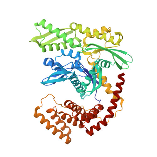
4J8F

Crystal structure of a fusion protein containing the NBD of Hsp70 and the middle domain of Hip


Experimental Data Snapshot

  • Method: X-RAY DIFFRACTION
  • Resolution: 2.70 Å
  • R-Value Free: 0.261 
  • R-Value Work: 0.194 
  • R-Value Observed: 0.197 

wwPDB Validation   3D Report Full Report


Ligand Structure Quality Assessment 


This is version 1.3 of the entry. See complete history


Literature

Structure and function of Hip, an attenuator of the Hsp70 chaperone cycle.

Li, Z.Hartl, F.U.Bracher, A.

(2013) Nat Struct Mol Biol 20: 929-935

  • DOI: https://doi.org/10.1038/nsmb.2608
  • Primary Citation of Related Structures:  
    4J8C, 4J8D, 4J8E, 4J8F

  • PubMed Abstract: 

    The Hsp70-interacting protein, Hip, cooperates with the chaperone Hsp70 in protein folding and prevention of aggregation. Hsp70 interacts with non-native protein substrates in an ATP-dependent reaction cycle regulated by J-domain proteins and nucleotide exchange factors (NEFs). Hip is thought to delay substrate release by slowing ADP dissociation from Hsp70. Here we present crystal structures of the dimerization domain and the tetratricopeptide repeat (TPR) domain of rat Hip. As shown in a cocrystal structure, the TPR core of Hip interacts with the Hsp70 ATPase domain through an extensive interface, to form a bracket that locks ADP in the binding cleft. Hip and NEF binding to Hsp70 are mutually exclusive, and thus Hip attenuates active cycling of Hsp70-substrate complexes. This mechanism explains how Hip enhances aggregation prevention by Hsp70 and facilitates transfer of specific proteins to downstream chaperones or the proteasome.


  • Organizational Affiliation

    Department of Cellular Biochemistry, Max Planck Institute of Biochemistry, Martinsried, Germany.


Macromolecules
Find similar proteins by:  (by identity cutoff)  |  3D Structure
Entity ID: 1
MoleculeChains Sequence LengthOrganismDetailsImage
Heat shock 70 kDa protein 1A/1B, Hsc70-interacting protein561Homo sapiensRattus norvegicus
This entity is chimeric
Mutation(s): 0 
Gene Names: HSPA1HSPA1AHSPA1BST13Fam10a1Hip
UniProt & NIH Common Fund Data Resources
Find proteins for P0DMV8 (Homo sapiens)
Explore P0DMV8 
Go to UniProtKB:  P0DMV8
PHAROS:  P0DMV8
GTEx:  ENSG00000204389 
Find proteins for P50503 (Rattus norvegicus)
Explore P50503 
Go to UniProtKB:  P50503
Entity Groups  
Sequence Clusters30% Identity50% Identity70% Identity90% Identity95% Identity100% Identity
UniProt GroupsP50503P0DMV8
Sequence Annotations
Expand
  • Reference Sequence
Small Molecules
Ligands 4 Unique
IDChains Name / Formula / InChI Key2D Diagram3D Interactions
ADP
Query on ADP

Download Ideal Coordinates CCD File 
B [auth A]ADENOSINE-5'-DIPHOSPHATE
C10 H15 N5 O10 P2
XTWYTFMLZFPYCI-KQYNXXCUSA-N
IOD
Query on IOD

Download Ideal Coordinates CCD File 
E [auth A]
F [auth A]
G [auth A]
H [auth A]
I [auth A]
E [auth A],
F [auth A],
G [auth A],
H [auth A],
I [auth A],
J [auth A],
K [auth A],
L [auth A],
M [auth A],
N [auth A],
O [auth A],
P [auth A],
Q [auth A],
R [auth A],
S [auth A],
T [auth A],
U [auth A]
IODIDE ION
I
XMBWDFGMSWQBCA-UHFFFAOYSA-M
PO4
Query on PO4

Download Ideal Coordinates CCD File 
D [auth A]PHOSPHATE ION
O4 P
NBIIXXVUZAFLBC-UHFFFAOYSA-K
MG
Query on MG

Download Ideal Coordinates CCD File 
C [auth A]MAGNESIUM ION
Mg
JLVVSXFLKOJNIY-UHFFFAOYSA-N
Experimental Data & Validation

Experimental Data

  • Method: X-RAY DIFFRACTION
  • Resolution: 2.70 Å
  • R-Value Free: 0.261 
  • R-Value Work: 0.194 
  • R-Value Observed: 0.197 
  • Space Group: P 41
Unit Cell:
Length ( Å )Angle ( ˚ )
a = 65.596α = 90
b = 65.596β = 90
c = 141.47γ = 90
Software Package:
Software NamePurpose
SCALAdata scaling
MOLREPphasing
REFMACrefinement
PDB_EXTRACTdata extraction
XDSdata scaling
XSCALEdata scaling

Structure Validation

View Full Validation Report


Ligand Structure Quality Assessment 


Entry History 

Deposition Data

Revision History  (Full details and data files)

  • Version 1.0: 2013-07-03
    Type: Initial release
  • Version 1.1: 2013-08-28
    Changes: Database references
  • Version 1.2: 2017-08-23
    Changes: Refinement description, Source and taxonomy
  • Version 1.3: 2024-02-28
    Changes: Data collection, Database references, Derived calculations