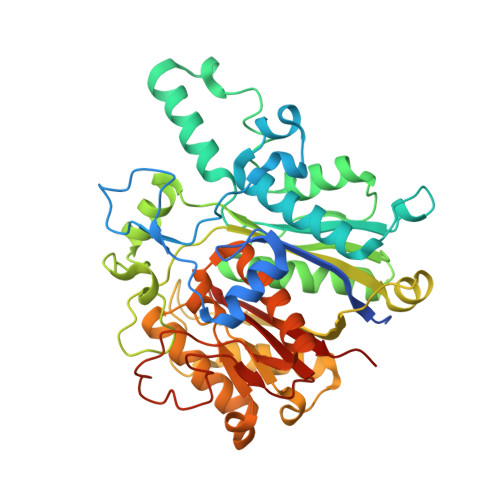
4JPF

Structure of wild type Pseudomonas aeruginosa FabF (KASII) in Complex with ligand


Experimental Data Snapshot

  • Method: X-RAY DIFFRACTION
  • Resolution: 1.67 Å
  • R-Value Free: 0.202 
  • R-Value Work: 0.169 
  • R-Value Observed: 0.171 

Starting Model: experimental
View more details

wwPDB Validation   3D Report Full Report


Ligand Structure Quality Assessment 


This is version 1.5 of the entry. See complete history


Literature

Structures of Pseudomonas aeruginosa beta-ketoacyl-(acyl-carrier-protein) synthase II (FabF) and a C164Q mutant provide templates for antibacterial drug discovery and identify a buried potassium ion and a ligand-binding site that is an artefact of the crystal form.

Baum, B.Lecker, L.S.Zoltner, M.Jaenicke, E.Schnell, R.Hunter, W.N.Brenk, R.

(2015) Acta Crystallogr Sect F Struct Biol Cryst Commun 71: 1020-1026

  • DOI: https://doi.org/10.1107/S2053230X15010614
  • Primary Citation of Related Structures:  
    4B7V, 4JB6, 4JPF

  • PubMed Abstract: 

    Bacterial infections remain a serious health concern, in particular causing life-threatening infections of hospitalized and immunocompromised patients. The situation is exacerbated by the rise in antibacterial drug resistance, and new treatments are urgently sought. In this endeavour, accurate structures of molecular targets can support early-stage drug discovery. Here, crystal structures, in three distinct forms, of recombinant Pseudomonas aeruginosa β-ketoacyl-(acyl-carrier-protein) synthase II (FabF) are presented. This enzyme, which is involved in fatty-acid biosynthesis, has been validated by genetic and chemical means as an antibiotic target in Gram-positive bacteria and represents a potential target in Gram-negative bacteria. The structures of apo FabF, of a C164Q mutant in which the binding site is altered to resemble the substrate-bound state and of a complex with 3-(benzoylamino)-2-hydroxybenzoic acid are reported. This compound mimics aspects of a known natural product inhibitor, platensimycin, and surprisingly was observed binding outside the active site, interacting with a symmetry-related molecule. An unusual feature is a completely buried potassium-binding site that was identified in all three structures. Comparisons suggest that this may represent a conserved structural feature of FabF relevant to fold stability. The new structures provide templates for structure-based ligand design and, together with the protocols and reagents, may underpin a target-based drug-discovery project for urgently needed antibacterials.


  • Organizational Affiliation

    Institut für Pharmazie und Biochemie, Johannes Gutenberg-Universität, Staudinger Weg 5, 55128 Mainz, Germany.


Macromolecules
Find similar proteins by:  (by identity cutoff)  |  3D Structure
Entity ID: 1
MoleculeChains Sequence LengthOrganismDetailsImage
3-oxoacyl-[acyl-carrier-protein] synthase 2436Pseudomonas aeruginosa PAO1Mutation(s): 0 
Gene Names: FabFfabF1PA2965
EC: 2.3.1.179
UniProt
Find proteins for G3XDA2 (Pseudomonas aeruginosa (strain ATCC 15692 / DSM 22644 / CIP 104116 / JCM 14847 / LMG 12228 / 1C / PRS 101 / PAO1))
Explore G3XDA2 
Go to UniProtKB:  G3XDA2
Entity Groups  
Sequence Clusters30% Identity50% Identity70% Identity90% Identity95% Identity100% Identity
UniProt GroupG3XDA2
Sequence Annotations
Expand
  • Reference Sequence
Small Molecules
Ligands 3 Unique
IDChains Name / Formula / InChI Key2D Diagram3D Interactions
1LR
Query on 1LR

Download Ideal Coordinates CCD File 
B [auth A]3-(benzoylamino)-2-hydroxybenzoic acid
C14 H11 N O4
ZIRLPQKKFAQFCV-UHFFFAOYSA-N
K
Query on K

Download Ideal Coordinates CCD File 
D [auth A]POTASSIUM ION
K
NPYPAHLBTDXSSS-UHFFFAOYSA-N
MG
Query on MG

Download Ideal Coordinates CCD File 
C [auth A]MAGNESIUM ION
Mg
JLVVSXFLKOJNIY-UHFFFAOYSA-N
Experimental Data & Validation

Experimental Data

  • Method: X-RAY DIFFRACTION
  • Resolution: 1.67 Å
  • R-Value Free: 0.202 
  • R-Value Work: 0.169 
  • R-Value Observed: 0.171 
  • Space Group: P 43 21 2
Unit Cell:
Length ( Å )Angle ( ˚ )
a = 71.6α = 90
b = 71.6β = 90
c = 140.03γ = 90
Software Package:
Software NamePurpose
MAR345dtbdata collection
PHASERphasing
REFMACrefinement
XDSdata reduction
XDSdata scaling

Structure Validation

View Full Validation Report


Ligand Structure Quality Assessment 


Entry History 

Deposition Data

Revision History  (Full details and data files)

  • Version 1.0: 2014-04-02
    Type: Initial release
  • Version 1.1: 2015-04-29
    Changes: Structure summary
  • Version 1.2: 2015-08-12
    Changes: Database references
  • Version 1.3: 2015-08-26
    Changes: Database references
  • Version 1.4: 2017-11-15
    Changes: Refinement description
  • Version 1.5: 2023-09-20
    Changes: Data collection, Database references, Derived calculations, Refinement description