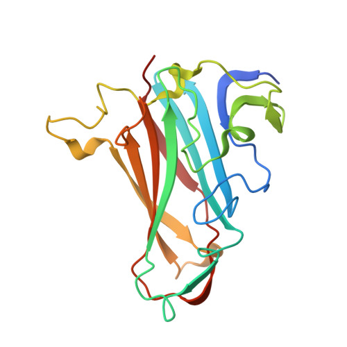
4K6T

Crystal structure of Ad37 fiber knob in complex with trivalent sialic acid inhibitor ME0385


Experimental Data Snapshot

  • Method: X-RAY DIFFRACTION
  • Resolution: 2.00 Å
  • R-Value Free: 0.200 
  • R-Value Work: 0.160 
  • R-Value Observed: 0.161 

Starting Model: experimental
View more details

wwPDB Validation   3D Report Full Report


Ligand Structure Quality Assessment 


This is version 1.6 of the entry. See complete history


Literature

Triazole linker-based trivalent sialic acid inhibitors of adenovirus type 37 infection of human corneal epithelial cells.

Caraballo, R.Saleeb, M.Bauer, J.Liaci, A.M.Chandra, N.Storm, R.J.Frangsmyr, L.Qian, W.Stehle, T.Arnberg, N.Elofsson, M.

(2015) Org Biomol Chem 13: 9194-9205

  • DOI: https://doi.org/10.1039/c5ob01025j
  • Primary Citation of Related Structures:  
    4K6T, 4K6U, 4K6V, 4K6W, 4XQA, 4XQB

  • PubMed Abstract: 

    Adenovirus type 37 (Ad37) is one of the principal agents responsible for epidemic keratoconjunctivitis (EKC), a severe ocular infection that remains without any available treatment. Recently, a trivalent sialic acid derivative (ME0322, Angew. Chem. Int. Ed., 2011, 50, 6519) was shown to function as a highly potent inhibitor of Ad37, efficiently preventing the attachment of the virion to the host cells and subsequent infection. Here, new trivalent sialic acid derivatives were designed, synthesized and their inhibitory properties against Ad37 infection of the human corneal epithelial cells were investigated. In comparison to ME0322, the best compound (17a) was found to be over three orders of magnitude more potent in a cell-attachment assay (IC50 = 1.4 nM) and about 140 times more potent in a cell-infection assay (IC50 = 2.9 nM). X-ray crystallographic analysis demonstrated a trivalent binding mode of all compounds to the Ad37 fiber knob. For the most potent compound ophthalmic toxicity in rabbits was investigated and it was concluded that repeated eye administration did not cause any adverse effects.


  • Organizational Affiliation

    Department of Chemistry, Umeå University, SE90187 Umeå, Sweden. mikael.elofsson@chem.umu.se.


Macromolecules
Find similar proteins by:  (by identity cutoff)  |  3D Structure
Entity ID: 1
MoleculeChains Sequence LengthOrganismDetailsImage
Fiber protein194Human adenovirus D37Mutation(s): 0 
Gene Names: L5
UniProt
Find proteins for Q64823 (Human adenovirus D37)
Explore Q64823 
Go to UniProtKB:  Q64823
Entity Groups  
Sequence Clusters30% Identity50% Identity70% Identity90% Identity95% Identity100% Identity
UniProt GroupQ64823
Sequence Annotations
Expand
  • Reference Sequence
Small Molecules
Ligands 9 Unique
IDChains Name / Formula / InChI Key2D Diagram3D Interactions
1P0
Query on 1P0

Download Ideal Coordinates CCD File 
Q [auth B]2,2',2''-[nitrilotris(methanediyl-1H-1,2,3-triazole-4,1-diyl)]triethanol
C15 H24 N10 O3
UBECCOXTPTYDBC-UHFFFAOYSA-N
SIA
Query on SIA

Download Ideal Coordinates CCD File 
AA [auth C]
DB [auth G]
G [auth A]
HA [auth E]
P [auth B]
AA [auth C],
DB [auth G],
G [auth A],
HA [auth E],
P [auth B],
TA [auth F]
N-acetyl-alpha-neuraminic acid
C11 H19 N O9
SQVRNKJHWKZAKO-YRMXFSIDSA-N
GOL
Query on GOL

Download Ideal Coordinates CCD File 
FB [auth G],
GB [auth G],
PA [auth E]
GLYCEROL
C3 H8 O3
PEDCQBHIVMGVHV-UHFFFAOYSA-N
ZN
Query on ZN

Download Ideal Coordinates CCD File 
AB [auth G]
BA [auth C]
BB [auth G]
CB [auth G]
FA [auth E]
AB [auth G],
BA [auth C],
BB [auth G],
CB [auth G],
FA [auth E],
GA [auth E],
H [auth A],
I [auth A],
J [auth A],
R [auth B],
RA [auth F],
S [auth B],
SA [auth F],
T [auth B]
ZINC ION
Zn
PTFCDOFLOPIGGS-UHFFFAOYSA-N
EDO
Query on EDO

Download Ideal Coordinates CCD File 
CA [auth C]
LA [auth E]
M [auth A]
MA [auth E]
N [auth A]
CA [auth C],
LA [auth E],
M [auth A],
MA [auth E],
N [auth A],
NA [auth E],
OA [auth E],
VA [auth F],
W [auth B],
WA [auth F],
X [auth B],
XA [auth F],
Y [auth B]
1,2-ETHANEDIOL
C2 H6 O2
LYCAIKOWRPUZTN-UHFFFAOYSA-N
ACT
Query on ACT

Download Ideal Coordinates CCD File 
EB [auth G]
IA [auth E]
JA [auth E]
K [auth A]
KA [auth E]
EB [auth G],
IA [auth E],
JA [auth E],
K [auth A],
KA [auth E],
L [auth A],
U [auth B],
UA [auth F],
V [auth B]
ACETATE ION
C2 H3 O2
QTBSBXVTEAMEQO-UHFFFAOYSA-M
CA
Query on CA

Download Ideal Coordinates CCD File 
HB [auth G]CALCIUM ION
Ca
BHPQYMZQTOCNFJ-UHFFFAOYSA-N
CL
Query on CL

Download Ideal Coordinates CCD File 
O [auth A]CHLORIDE ION
Cl
VEXZGXHMUGYJMC-UHFFFAOYSA-M
MG
Query on MG

Download Ideal Coordinates CCD File 
DA [auth C]
EA [auth C]
QA [auth E]
YA [auth F]
Z [auth B]
DA [auth C],
EA [auth C],
QA [auth E],
YA [auth F],
Z [auth B],
ZA [auth F]
MAGNESIUM ION
Mg
JLVVSXFLKOJNIY-UHFFFAOYSA-N
Experimental Data & Validation

Experimental Data

  • Method: X-RAY DIFFRACTION
  • Resolution: 2.00 Å
  • R-Value Free: 0.200 
  • R-Value Work: 0.160 
  • R-Value Observed: 0.161 
  • Space Group: P 1 21 1
Unit Cell:
Length ( Å )Angle ( ˚ )
a = 74.94α = 90
b = 67.18β = 96.81
c = 121.43γ = 90
Software Package:
Software NamePurpose
RemDAqdata collection
PHASERphasing
REFMACrefinement
XDSdata reduction
XSCALEdata scaling

Structure Validation

View Full Validation Report


Ligand Structure Quality Assessment 


Entry History 

Deposition Data

Revision History  (Full details and data files)

  • Version 1.0: 2014-10-22
    Type: Initial release
  • Version 1.1: 2015-07-29
    Changes: Database references
  • Version 1.2: 2015-09-09
    Changes: Database references
  • Version 1.3: 2017-11-15
    Changes: Refinement description
  • Version 1.4: 2020-07-29
    Type: Remediation
    Reason: Carbohydrate remediation
    Changes: Data collection, Database references, Derived calculations, Structure summary
  • Version 1.5: 2024-02-28
    Changes: Data collection, Database references, Structure summary
  • Version 1.6: 2024-04-03
    Changes: Refinement description