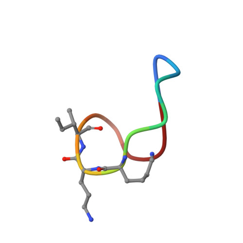
4K7T

Structure of the ternary complex of bacitracin, zinc, and geranyl-pyrophosphate


Experimental Data Snapshot

  • Method: X-RAY DIFFRACTION
  • Resolution: 1.10 Å
  • R-Value Free: 0.149 
  • R-Value Work: 0.127 
  • R-Value Observed: 0.128 

wwPDB Validation   3D Report Full Report


Ligand Structure Quality Assessment 


This is version 1.3 of the entry. See complete history


Literature

High-resolution crystal structure reveals molecular details of target recognition by bacitracin.

Economou, N.J.Cocklin, S.Loll, P.J.

(2013) Proc Natl Acad Sci U S A 110: 14207-14212

  • DOI: https://doi.org/10.1073/pnas.1308268110
  • Primary Citation of Related Structures:  
    4K7T

  • PubMed Abstract: 

    Bacitracin is a metalloantibiotic agent that is widely used as a medicine and feed additive. It interferes with bacterial cell-wall biosynthesis by binding undecaprenyl-pyrophosphate, a lipid carrier that serves as a critical intermediate in cell wall production. Despite bacitracin's broad use, the molecular details of its target recognition have not been elucidated. Here we report a crystal structure for the ternary complex of bacitracin A, zinc, and a geranyl-pyrophosphate ligand at a resolution of 1.1 Å. The antibiotic forms a compact structure that completely envelopes the ligand's pyrophosphate group, together with flanking zinc and sodium ions. The complex adopts a highly amphipathic conformation that offers clues to antibiotic function in the context of bacterial membranes. Bacitracin's efficient sequestration of its target represents a previously unseen mode for the recognition of lipid pyrophosphates, and suggests new directions for the design of next-generation antimicrobial agents.


  • Organizational Affiliation

    Department of Biochemistry and Molecular Biology, Drexel University College of Medicine, Philadelphia, PA 19102, USA.


Macromolecules

Find similar proteins by:  Sequence   |   3D Structure  

Entity ID: 1
MoleculeChains Sequence LengthOrganismDetailsImage
bacitracin A212Bacillus subtilisMutation(s): 0 
Entity Groups  
Sequence Clusters30% Identity50% Identity70% Identity90% Identity95% Identity100% Identity
Sequence Annotations
Expand
  • Reference Sequence
Biologically Interesting Molecules (External Reference) 1 Unique
Experimental Data & Validation

Experimental Data

  • Method: X-RAY DIFFRACTION
  • Resolution: 1.10 Å
  • R-Value Free: 0.149 
  • R-Value Work: 0.127 
  • R-Value Observed: 0.128 
  • Space Group: P 62
Unit Cell:
Length ( Å )Angle ( ˚ )
a = 35.98α = 90
b = 35.98β = 90
c = 16.12γ = 120
Software Package:
Software NamePurpose
XSCALEdata scaling
PHENIXrefinement
PDB_EXTRACTdata extraction
ADSCdata collection
XDSdata reduction
PHENIXphasing

Structure Validation

View Full Validation Report


Ligand Structure Quality Assessment 


Entry History 

Deposition Data

Revision History  (Full details and data files)

  • Version 1.0: 2013-08-14
    Type: Initial release
  • Version 1.1: 2013-09-18
    Changes: Database references
  • Version 1.2: 2013-10-16
    Changes: Derived calculations
  • Version 1.3: 2024-10-16
    Changes: Data collection, Database references, Derived calculations, Structure summary