4KIK | pdb_00004kik

Human IkB kinase beta


Experimental Data Snapshot

  • Method: X-RAY DIFFRACTION
  • Resolution: 2.83 Å
  • R-Value Free: 
    0.236 (Depositor), 0.247 (DCC) 
  • R-Value Work: 
    0.184 (Depositor), 0.192 (DCC) 
  • R-Value Observed: 
    0.186 (Depositor) 

wwPDB Validation 3D Report Full Report

Validation slider image for 4KIK

Ligand Structure Quality Assessment 


This is version 1.2 of the entry. See complete history

Literature

Crystal Structure of a Human I kappa B Kinase beta Asymmetric Dimer.

Liu, S.Misquitta, Y.R.Olland, A.Johnson, M.A.Kelleher, K.S.Kriz, R.Lin, L.L.Stahl, M.Mosyak, L.

(2013) J Biological Chem 288: 22758-22767

  • DOI: https://doi.org/10.1074/jbc.M113.482596
  • Primary Citation Related Structures: 
    4KIK

  • PubMed Abstract: 

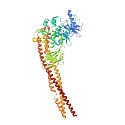
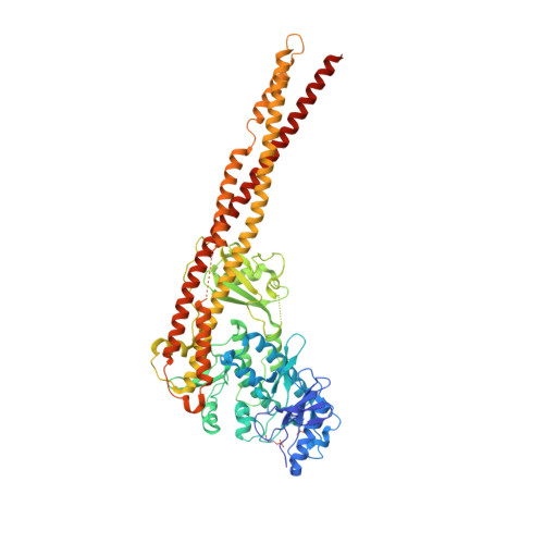
    Phosphorylation of inhibitor of nuclear transcription factor κB (IκB) by IκB kinase (IKK) triggers the degradation of IκB and migration of cytoplasmic κB to the nucleus where it promotes the transcription of its target genes. Activation of IKK is achieved by phosphorylation of its main subunit, IKKβ, at the activation loop sites. Here, we report the 2.8 Å resolution crystal structure of human IKKβ (hIKKβ), which is partially phosphorylated and bound to the staurosporine analog K252a. The hIKKβ protomer adopts a trimodular structure that closely resembles that from Xenopus laevis (xIKKβ): an N-terminal kinase domain (KD), a central ubiquitin-like domain (ULD), and a C-terminal scaffold/dimerization domain (SDD). Although hIKKβ and xIKKβ utilize a similar dimerization mode, their overall geometries are distinct. In contrast to the structure resembling closed shears reported previously for xIKKβ, hIKKβ exists as an open asymmetric dimer in which the two KDs are further apart, with one in an active and the other in an inactive conformation. Dimer interactions are limited to the C-terminal six-helix bundle that acts as a hinge between the two subunits. The observed domain movements in the structures of IKKβ may represent trans-phosphorylation steps that accompany IKKβ activation.


  • Organizational Affiliation
    • Structural Biology and Biophysics Group, Pfizer Worldwide Research, Groton, Connecticut 06340, USA. shenping.liu@pfizer.com

Macromolecule Content 

  • Total Structure Weight: 157.52 kDa 
  • Atom Count: 10,602 
  • Modeled Residue Count: 1,269 
  • Deposited Residue Count: 1,354 
  • Unique protein chains: 2

Macromolecules

Find similar proteins by:|  3D Structure
Entity ID: 1
MoleculeChains  Sequence LengthOrganismDetailsImage
Inhibitor of nuclear factor kappa-B kinase subunit beta677Homo sapiensMutation(s): 0 
Gene Names: IKBKBIKKB
EC: 2.7.11.10 (PDB Primary Data), 2.7.11.1 (UniProt)
UniProt & NIH Common Fund Data Resources
Find proteins for O14920 (Homo sapiens)
Explore O14920 
Go to UniProtKB:  O14920
PHAROS:  O14920
GTEx:  ENSG00000104365 
Entity Groups
Sequence Clusters30% Identity50% Identity70% Identity90% Identity95% Identity100% Identity
UniProt GroupO14920
Sequence Annotations
Expand
Reference Sequence
Find similar proteins by:|  3D Structure
Entity ID: 2
MoleculeChains  Sequence LengthOrganismDetailsImage
Inhibitor of nuclear factor kappa-B kinase subunit beta677Homo sapiensMutation(s): 0 
Gene Names: IKBKBIKKB
EC: 2.7.11.10 (PDB Primary Data), 2.7.11.1 (UniProt)
UniProt & NIH Common Fund Data Resources
Find proteins for O14920 (Homo sapiens)
Explore O14920 
Go to UniProtKB:  O14920
PHAROS:  O14920
GTEx:  ENSG00000104365 
Entity Groups
Sequence Clusters30% Identity50% Identity70% Identity90% Identity95% Identity100% Identity
UniProt GroupO14920
Sequence Annotations
Expand
Reference Sequence

Small Molecules

Modified Residues  1 Unique
IDChains TypeFormula2D DiagramParent
SEP
Query on SEP
A
L-PEPTIDE LINKINGC3 H8 N O6 PSER

Experimental Data & Validation

Experimental Data

  • Method: X-RAY DIFFRACTION
  • Resolution: 2.83 Å
  • R-Value Free:  0.236 (Depositor), 0.247 (DCC) 
  • R-Value Work:  0.184 (Depositor), 0.192 (DCC) 
  • R-Value Observed: 0.186 (Depositor) 
Space Group: P 1 21 1
Unit Cell:
Length ( Å )Angle ( ˚ )
a = 110.954α = 90
b = 68.689β = 107.03
c = 107.401γ = 90
Software Package:
Software NamePurpose
BUSTERrefinement

Structure Validation

View Full Validation Report


Ligand Structure Quality Assessment 


Entry History 

Deposition Data

Revision History  (Full details and data files)

  • Version 1.0: 2013-06-26
    Type: Initial release
  • Version 1.1: 2013-08-28
    Changes: Database references
  • Version 1.2: 2024-10-30
    Changes: Data collection, Database references, Derived calculations, Structure summary