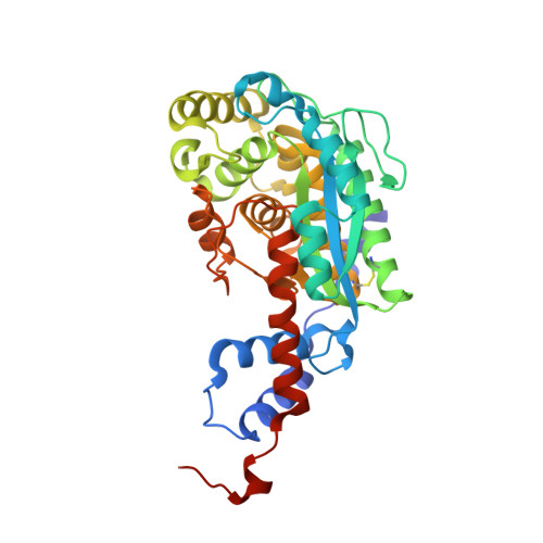
Dissecting cobamide diversity through structural and functional analyses of the base-activating CobT enzyme of Salmonella enterica.
Chan, C.H., Newmister, S.A., Talyor, K., Claas, K.R., Rayment, I., Escalante-Semerena, J.C.(2014) Biochim Biophys Acta 1840: 464-475
- PubMed: 24121107
- DOI: https://doi.org/10.1016/j.bbagen.2013.09.038
- Primary Citation of Related Structures:
4KQF, 4KQG, 4KQH, 4KQI, 4KQJ, 4KQK - PubMed Abstract:
Cobamide diversity arises from the nature of the nucleotide base. Nicotinate mononucleotide (NaMN):base phosphoribosyltransferases (CobT) synthesize α-linked riboside monophosphates from diverse nucleotide base substrates (e.g., benzimidazoles, purines, phenolics) that are incorporated into cobamides. Structural investigations of two members of the CobT family of enzymes in complex with various substrate bases as well as in vivo and vitro activity analyses of enzyme variants were performed to elucidate the roles of key amino acid residues important for substrate recognition. Results of in vitro and in vivo studies of active-site variants of the Salmonella enterica CobT (SeCobT) enzyme suggest that a catalytic base may not be required for catalysis. This idea is supported by the analyses of crystal structures that show that two glutamate residues function primarily to maintain an active conformation of the enzyme. In light of these findings, we propose that proper positioning of the substrates in the active site triggers the attack at the C1 ribose of NaMN. Whether or not a catalytic base is needed for function is discussed within the framework of the in vitro analysis of the enzyme activity. Additionally, structure-guided site-directed mutagenesis of SeCobT broadened its substrate specificity to include phenolic bases, revealing likely evolutionary changes needed to increase cobamide diversity, and further supporting the proposed mechanism for the phosphoribosylation of phenolic substrates. Results of this study uncover key residues in the CobT enzyme that contribute to the diversity of cobamides in nature.
- Department of Bacteriology, University of Wisconsin, Madison, WI, USA.
Organizational Affiliation:


