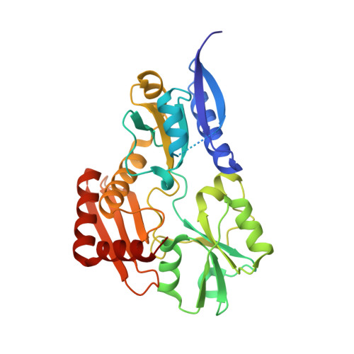
4MLV

Crystal Structure of Bacillus megaterium porphobilinogen deaminase


Experimental Data Snapshot

  • Method: X-RAY DIFFRACTION
  • Resolution: 1.46 Å
  • R-Value Free: 0.186 
  • R-Value Work: 0.141 
  • R-Value Observed: 0.143 

Starting Model: experimental
View more details

wwPDB Validation   3D Report Full Report


Ligand Structure Quality Assessment 


This is version 1.1 of the entry. See complete history


Literature

Structural evidence for the partially oxidized dipyrromethene and dipyrromethanone forms of the cofactor of porphobilinogen deaminase: structures of the Bacillus megaterium enzyme at near-atomic resolution.

Azim, N.Deery, E.Warren, M.J.Wolfenden, B.A.Erskine, P.Cooper, J.B.Coker, A.Wood, S.P.Akhtar, M.

(2014) Acta Crystallogr D Biol Crystallogr 70: 744-751

  • DOI: https://doi.org/10.1107/S139900471303294X
  • Primary Citation of Related Structures:  
    4MLQ, 4MLV

  • PubMed Abstract: 

    The enzyme porphobilinogen deaminase (PBGD; hydroxymethylbilane synthase; EC 2.5.1.61) catalyses an early step of the tetrapyrrole-biosynthesis pathway in which four molecules of the monopyrrole porphobilinogen are condensed to form a linear tetrapyrrole. The enzyme possesses a dipyrromethane cofactor, which is covalently linked by a thioether bridge to an invariant cysteine residue (Cys241 in the Bacillus megaterium enzyme). The cofactor is extended during the reaction by the sequential addition of the four substrate molecules, which are released as a linear tetrapyrrole product. Expression in Escherichia coli of a His-tagged form of B. megaterium PBGD has permitted the X-ray analysis of the enzyme from this species at high resolution, showing that the cofactor becomes progressively oxidized to the dipyrromethene and dipyrromethanone forms. In previously solved PBGD structures, the oxidized cofactor is in the dipyromethenone form, in which both pyrrole rings are approximately coplanar. In contrast, the oxidized cofactor in the B. megaterium enzyme appears to be in the dipyrromethanone form, in which the C atom at the bridging α-position of the outer pyrrole ring is very clearly in a tetrahedral configuration. It is suggested that the pink colour of the freshly purified protein is owing to the presence of the dipyrromethene form of the cofactor which, in the structure reported here, adopts the same conformation as the fully reduced dipyrromethane form.


  • Organizational Affiliation

    School of Biological Sciences, University of Punjab, New Campus, Lahore-54590, Pakistan.


Macromolecules
Find similar proteins by:  (by identity cutoff)  |  3D Structure
Entity ID: 1
MoleculeChains Sequence LengthOrganismDetailsImage
Porphobilinogen deaminase312Priestia megateriumMutation(s): 0 
Gene Names: hemC
EC: 2.5.1.61
UniProt
Find proteins for Q8GCA8 (Priestia megaterium)
Explore Q8GCA8 
Go to UniProtKB:  Q8GCA8
Entity Groups  
Sequence Clusters30% Identity50% Identity70% Identity90% Identity95% Identity100% Identity
UniProt GroupQ8GCA8
Sequence Annotations
Expand
  • Reference Sequence
Small Molecules
Ligands 3 Unique
IDChains Name / Formula / InChI Key2D Diagram3D Interactions
29P
Query on 29P

Download Ideal Coordinates CCD File 
C [auth A]3-[(5S)-5-{[3-(2-carboxyethyl)-4-(carboxymethyl)-5-methyl-1H-pyrrol-2-yl]methyl}-4-(carboxymethyl)-2-oxo-2,5-dihydro-1H-pyrrol-3-yl]propanoic acid
C20 H24 N2 O9
DHEOBTWDCMSDOQ-HNNXBMFYSA-N
DPM
Query on DPM

Download Ideal Coordinates CCD File 
B [auth A]3-[5-{[3-(2-carboxyethyl)-4-(carboxymethyl)-5-methyl-1H-pyrrol-2-yl]methyl}-4-(carboxymethyl)-1H-pyrrol-3-yl]propanoic acid
C20 H24 N2 O8
LCAXMKQKEYTFDM-UHFFFAOYSA-N
ACY
Query on ACY

Download Ideal Coordinates CCD File 
D [auth A]ACETIC ACID
C2 H4 O2
QTBSBXVTEAMEQO-UHFFFAOYSA-N
Experimental Data & Validation

Experimental Data

  • Method: X-RAY DIFFRACTION
  • Resolution: 1.46 Å
  • R-Value Free: 0.186 
  • R-Value Work: 0.141 
  • R-Value Observed: 0.143 
  • Space Group: P 21 21 21
Unit Cell:
Length ( Å )Angle ( ˚ )
a = 53.32α = 90
b = 65.78β = 90
c = 97.21γ = 90
Software Package:
Software NamePurpose
GDAdata collection
MOLREPphasing
REFMACrefinement
MOSFLMdata reduction
SCALAdata scaling

Structure Validation

View Full Validation Report


Ligand Structure Quality Assessment 


Entry History 

Deposition Data

Revision History  (Full details and data files)

  • Version 1.0: 2014-04-02
    Type: Initial release
  • Version 1.1: 2023-09-20
    Changes: Data collection, Database references, Derived calculations, Refinement description