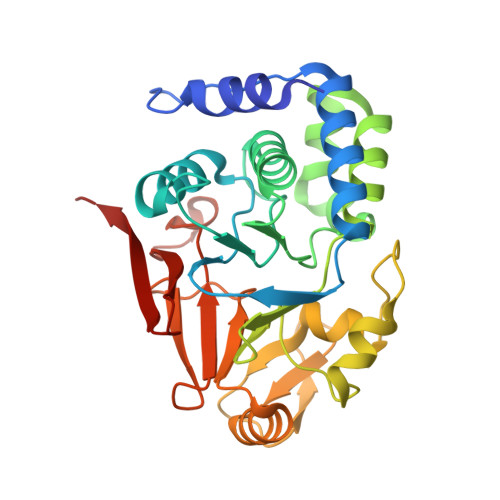
4MOY

Structure of a second nuclear PP1 Holoenzyme, crystal form 1


Experimental Data Snapshot

  • Method: X-RAY DIFFRACTION
  • Resolution: 2.20 Å
  • R-Value Free: 0.185 
  • R-Value Work: 0.151 
  • R-Value Observed: 0.153 

wwPDB Validation   3D Report Full Report


This is version 1.2 of the entry. See complete history


Literature

Understanding the antagonism of retinoblastoma protein dephosphorylation by PNUTS provides insights into the PP1 regulatory code.

Choy, M.S.Hieke, M.Kumar, G.S.Lewis, G.R.Gonzalez-Dewhitt, K.R.Kessler, R.P.Stein, B.J.Hessenberger, M.Nairn, A.C.Peti, W.Page, R.

(2014) Proc Natl Acad Sci U S A 111: 4097-4102

  • DOI: https://doi.org/10.1073/pnas.1317395111
  • Primary Citation of Related Structures:  
    4MOV, 4MOY, 4MP0

  • PubMed Abstract: 

    The serine/threonine protein phosphatase 1 (PP1) dephosphorylates hundreds of key biological targets by associating with nearly 200 regulatory proteins to form highly specific holoenzymes. However, how these proteins direct PP1 specificity and the ability to predict how these PP1 interacting proteins bind PP1 from sequence alone is still missing. PP1 nuclear targeting subunit (PNUTS) is a PP1 targeting protein that, with PP1, plays a central role in the nucleus, where it regulates chromatin decondensation, RNA processing, and the phosphorylation state of fundamental cell cycle proteins, including the retinoblastoma protein (Rb), p53, and MDM2. The molecular function of PNUTS in these processes is completely unknown. Here, we show that PNUTS, which is intrinsically disordered in its free form, interacts strongly with PP1 in a highly extended manner. Unexpectedly, PNUTS blocks one of PP1's substrate binding grooves while leaving the active site accessible. This interaction site, which we have named the arginine site, allowed us to define unique PP1 binding motifs, which advances our ability to predict how more than a quarter of the known PP1 regulators bind PP1. Additionally, the structure shows how PNUTS inhibits the PP1-mediated dephosphorylation of critical substrates, especially Rb, by blocking their binding sites on PP1, insights that are providing strategies for selectively enhancing Rb activity.


  • Organizational Affiliation

    Departments of Molecular Pharmacology, Physiology, and Biotechnology, Molecular Biology, Cell Biology, and Biochemistry, and Chemistry, Brown University, Providence, RI 02912.


Macromolecules
Find similar proteins by:  (by identity cutoff)  |  3D Structure
Entity ID: 1
MoleculeChains Sequence LengthOrganismDetailsImage
Serine/threonine-protein phosphatase PP1-alpha catalytic subunit299Homo sapiensMutation(s): 0 
Gene Names: PPP1CAPPP1A
EC: 3.1.3.16
UniProt & NIH Common Fund Data Resources
Find proteins for P62136 (Homo sapiens)
Explore P62136 
Go to UniProtKB:  P62136
PHAROS:  P62136
GTEx:  ENSG00000172531 
Entity Groups  
Sequence Clusters30% Identity50% Identity70% Identity90% Identity95% Identity100% Identity
UniProt GroupP62136
Sequence Annotations
Expand
  • Reference Sequence
Find similar proteins by:  (by identity cutoff)  |  3D Structure
Entity ID: 2
MoleculeChains Sequence LengthOrganismDetailsImage
Serine/threonine-protein phosphatase 1 regulatory subunit 1044Rattus norvegicusMutation(s): 0 
Gene Names: Ppp1r10Cat53Pnuts
UniProt
Find proteins for O55000 (Rattus norvegicus)
Explore O55000 
Go to UniProtKB:  O55000
Entity Groups  
Sequence Clusters30% Identity50% Identity70% Identity90% Identity95% Identity100% Identity
UniProt GroupO55000
Sequence Annotations
Expand
  • Reference Sequence
Small Molecules
Ligands 4 Unique
IDChains Name / Formula / InChI Key2D Diagram3D Interactions
PO4
Query on PO4

Download Ideal Coordinates CCD File 
F [auth A]PHOSPHATE ION
O4 P
NBIIXXVUZAFLBC-UHFFFAOYSA-K
GOL
Query on GOL

Download Ideal Coordinates CCD File 
G [auth A],
H [auth A]
GLYCEROL
C3 H8 O3
PEDCQBHIVMGVHV-UHFFFAOYSA-N
MN
Query on MN

Download Ideal Coordinates CCD File 
C [auth A],
D [auth A]
MANGANESE (II) ION
Mn
WAEMQWOKJMHJLA-UHFFFAOYSA-N
CL
Query on CL

Download Ideal Coordinates CCD File 
E [auth A]CHLORIDE ION
Cl
VEXZGXHMUGYJMC-UHFFFAOYSA-M
Experimental Data & Validation

Experimental Data

  • Method: X-RAY DIFFRACTION
  • Resolution: 2.20 Å
  • R-Value Free: 0.185 
  • R-Value Work: 0.151 
  • R-Value Observed: 0.153 
  • Space Group: P 32 2 1
Unit Cell:
Length ( Å )Angle ( ˚ )
a = 130.788α = 90
b = 130.788β = 90
c = 47.725γ = 120
Software Package:
Software NamePurpose
CBASSdata collection
PHENIXmodel building
PHENIXrefinement
HKL-2000data reduction
HKL-2000data scaling
PHENIXphasing

Structure Validation

View Full Validation Report


Entry History 

Deposition Data

Revision History  (Full details and data files)

  • Version 1.0: 2014-03-26
    Type: Initial release
  • Version 1.1: 2014-04-23
    Changes: Database references
  • Version 1.2: 2024-02-28
    Changes: Data collection, Database references, Derived calculations