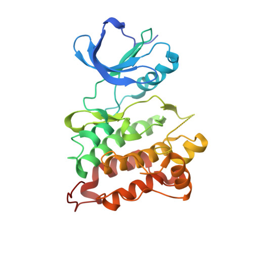
4MXO

human Src kinase bound to kinase inhibitor bosutinib


Experimental Data Snapshot

  • Method: X-RAY DIFFRACTION
  • Resolution: 2.10 Å
  • R-Value Free: 0.254 
  • R-Value Work: 0.210 
  • R-Value Observed: 0.212 

wwPDB Validation   3D Report Full Report


Ligand Structure Quality Assessment 


This is version 1.2 of the entry. See complete history


Literature

A conserved water-mediated hydrogen bond network defines bosutinib's kinase selectivity.

Levinson, N.M.Boxer, S.G.

(2014) Nat Chem Biol 10: 127-132

  • DOI: https://doi.org/10.1038/nchembio.1404
  • Primary Citation of Related Structures:  
    4MXO, 4MXX, 4MXY, 4MXZ

  • PubMed Abstract: 

    Kinase inhibitors are important cancer drugs, but they tend to display limited target specificity, and their target profiles are often challenging to rationalize in terms of molecular mechanism. Here we report that the clinical kinase inhibitor bosutinib recognizes its kinase targets by engaging a pair of conserved structured water molecules in the active site and that many other kinase inhibitors share a similar recognition mechanism. Using the nitrile group of bosutinib as an infrared probe, we show that the gatekeeper residue and one other position in the ATP-binding site control access of the drug to the structured water molecules and that the amino acids found at these positions account for the kinome-wide target spectrum of the drug. Our work highlights the importance of structured water molecules for inhibitor recognition, reveals a new role for the kinase gatekeeper and showcases an effective approach for elucidating the molecular origins of selectivity patterns.


  • Organizational Affiliation

    Department of Chemistry, Stanford University, Stanford, California, USA.


Macromolecules
Find similar proteins by:  (by identity cutoff)  |  3D Structure
Entity ID: 1
MoleculeChains Sequence LengthOrganismDetailsImage
Proto-oncogene tyrosine-protein kinase Src
A, B
286Homo sapiensMutation(s): 0 
Gene Names: SRCSRC1
EC: 2.7.10.2
UniProt & NIH Common Fund Data Resources
Find proteins for P12931 (Homo sapiens)
Explore P12931 
Go to UniProtKB:  P12931
PHAROS:  P12931
GTEx:  ENSG00000197122 
Entity Groups  
Sequence Clusters30% Identity50% Identity70% Identity90% Identity95% Identity100% Identity
UniProt GroupP12931
Sequence Annotations
Expand
  • Reference Sequence
Small Molecules
Ligands 1 Unique
IDChains Name / Formula / InChI Key2D Diagram3D Interactions
DB8
Query on DB8

Download Ideal Coordinates CCD File 
C [auth A],
D [auth B]
4-[(2,4-dichloro-5-methoxyphenyl)amino]-6-methoxy-7-[3-(4-methylpiperazin-1-yl)propoxy]quinoline-3-carbonitrile
C26 H29 Cl2 N5 O3
UBPYILGKFZZVDX-UHFFFAOYSA-N
Experimental Data & Validation

Experimental Data

  • Method: X-RAY DIFFRACTION
  • Resolution: 2.10 Å
  • R-Value Free: 0.254 
  • R-Value Work: 0.210 
  • R-Value Observed: 0.212 
  • Space Group: P 1
Unit Cell:
Length ( Å )Angle ( ˚ )
a = 41.87α = 79.53
b = 63.185β = 89.31
c = 73.665γ = 90.05
Software Package:
Software NamePurpose
Blu-Icedata collection
PHASERphasing
PHENIXrefinement
MOSFLMdata reduction
SCALAdata scaling

Structure Validation

View Full Validation Report


Ligand Structure Quality Assessment 


Entry History 

Deposition Data

Revision History  (Full details and data files)

  • Version 1.0: 2013-12-04
    Type: Initial release
  • Version 1.1: 2014-02-05
    Changes: Database references
  • Version 1.2: 2024-02-28
    Changes: Data collection, Database references, Derived calculations