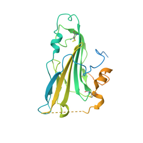
Crystal structure of herpes simplex virus 2 gD bound to nectin-1 reveals a conserved mode of receptor recognition.
Lu, G., Zhang, N., Qi, J., Li, Y., Chen, Z., Zheng, C., Gao, G.F., Yan, J.(2014) J Virol 88: 13678-13688
- PubMed: 25231300
- DOI: https://doi.org/10.1128/JVI.01906-14
- Primary Citation of Related Structures:
4MYV, 4MYW - PubMed Abstract:
Herpes simplex virus 1 (HSV-1) and HSV-2 are among the most prevalent human pathogens. Both viruses can recognize, via the surface envelope glycoprotein D (gD), human nectin-1 as a functional receptor. Previous studies have successfully elucidated the molecular basis of the binding between HSV-1 gD and nectin-1 by cocrystallography. Despite a high sequence identity between HSV-1 and HSV-2 gDs, the atomic intermolecule details for the HSV-2-gD/nectin-1 interaction remain elusive. Here, we report the crystal structures of both the unbound and the nectin-1-bound HSV-2 gDs. The free-gD structure expectedly comprises an IgV-like core and the surface-exposed terminal extensions as observed in its HSV-1 counterpart but lacks traceable electron densities for a large portion of the terminal elements. These terminal residues were clearly traced in the complex structure as a definitive loop in the N terminus and an α-helix in the C terminus, thereby showing a conserved nectin-1-binding mode as reported for HSV-1 gD. The interface residues in nectin-1 were further mutated and tested for the gD interaction by surface plasmon resonance. The resultant binding patterns were similar for HSV-1 and HSV-2 gDs, further supporting a homologous receptor-binding basis by the two viruses for nectin-1. These data, together with a cell-based fusion assay showing a cross-inhibition of the gD/nectin-1-mediated cell-cell fusion by soluble HSV-1 and HSV-2 gDs, provided solid structural and functional evidence that HSV-1 and HSV-2 recognize nectin-1 via the same binding mode. Finally, we also demonstrated that nectin-1 I80 is an important residue involved in gD interaction. Despite intensified studies, a detailed picture of the molecular features in the HSV-2-gD/nectin-1 interaction remains unavailable. Previous work focused on HSV-1 gD, which folds into an IgV-like core with large terminal extensions and utilizes the extension elements to engage nectin-1. Here, we report the crystal structures of HSV-2 gD in both the free and the nectin-1-bound forms. The atomic intermolecule details for HSV-2-gD/nectin-1 interaction are clearly presented. The observed binding mode is identical to that reported for its HSV-1 counterpart. This structural observation was further supported by our comparative functional assays showing that nectin-1 mutations similarly affect the ligand-receptor interaction of both virus gDs. Taken together, we provide comprehensive structural and functional data demonstrating a conserved receptor-binding mode between HSV-1 and HSV-2 for nectin-1. Our results also indicate that the tropism difference between the two viruses likely arises from aspects other than the gD/nectin-1 binding features.
- CAS Key Laboratory of Pathogenic Microbiology and Immunology, Institute of Microbiology, Chinese Academy of Sciences, Beijing, China.
Organizational Affiliation:


