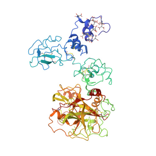
4O03

Crystal structure of Ca2+ bound prothrombin deletion mutant residues 146-167


Experimental Data Snapshot

  • Method: X-RAY DIFFRACTION
  • Resolution: 3.38 Å
  • R-Value Free: 0.279 
  • R-Value Work: 0.233 
  • R-Value Observed: 0.235 

Starting Model: experimental
View more details

wwPDB Validation   3D Report Full Report


Ligand Structure Quality Assessment 


This is version 1.5 of the entry. See complete history


Literature

The linker connecting the two kringles plays a key role in prothrombin activation.

Pozzi, N.Chen, Z.Pelc, L.A.Shropshire, D.B.Di Cera, E.

(2014) Proc Natl Acad Sci U S A 111: 7630-7635

  • DOI: https://doi.org/10.1073/pnas.1403779111
  • Primary Citation of Related Structures:  
    4NZQ, 4O03

  • PubMed Abstract: 

    The zymogen prothrombin is proteolytically converted by factor Xa to the active protease thrombin in a reaction that is accelerated >3,000-fold by cofactor Va. This physiologically important effect is paradigmatic of analogous cofactor-dependent reactions in the coagulation and complement cascades, but its structural determinants remain poorly understood. Prothrombin has three linkers connecting the N-terminal Gla domain to kringle-1 (Lnk1), the two kringles (Lnk2), and kringle-2 to the C-terminal protease domain (Lnk3). Recent developments indicate that the linkers, and particularly Lnk2, confer on the zymogen significant flexibility in solution and enable prothrombin to sample alternative conformations. The role of this flexibility in the context of prothrombin activation was tested with several deletions. Removal of Lnk2 in almost its entirety (ProTΔ146-167) drastically reduces the enhancement of thrombin generation by cofactor Va from >3,000-fold to 60-fold because of a significant increase in the rate of activation in the absence of cofactor. Deletion of Lnk2 mimics the action of cofactor Va and offers insights into how prothrombin is activated at the molecular level. The crystal structure of ProTΔ146-167 reveals a contorted architecture where the domains are not vertically stacked, kringle-1 comes within 9 Å of the protease domain, and the Gla-domain primed for membrane binding comes in contact with kringle-2. These findings broaden our molecular understanding of a key reaction of the blood coagulation cascade where cofactor Va enhances activation of prothrombin by factor Xa by compressing Lnk2 and morphing prothrombin into a conformation similar to the structure of ProTΔ146-167.


  • Organizational Affiliation

    Edward A. Doisy Department of Biochemistry and Molecular Biology, Saint Louis University School of Medicine, St. Louis, MO 63104.


Macromolecules
Find similar proteins by:  (by identity cutoff)  |  3D Structure
Entity ID: 1
MoleculeChains Sequence LengthOrganismDetailsImage
Prothrombin557Homo sapiensMutation(s): 0 
Gene Names: F2
EC: 3.4.21.5
Membrane Entity: Yes 
UniProt & NIH Common Fund Data Resources
Find proteins for P00734 (Homo sapiens)
Explore P00734 
Go to UniProtKB:  P00734
PHAROS:  P00734
GTEx:  ENSG00000180210 
Entity Groups  
Sequence Clusters30% Identity50% Identity70% Identity90% Identity95% Identity100% Identity
UniProt GroupP00734
Glycosylation
Glycosylation Sites: 3Go to GlyGen: P00734-1
Sequence Annotations
Expand
  • Reference Sequence
Small Molecules
Ligands 2 Unique
IDChains Name / Formula / InChI Key2D Diagram3D Interactions
NAG
Query on NAG

Download Ideal Coordinates CCD File 
G [auth A],
H [auth A],
I [auth A]
2-acetamido-2-deoxy-beta-D-glucopyranose
C8 H15 N O6
OVRNDRQMDRJTHS-FMDGEEDCSA-N
CA
Query on CA

Download Ideal Coordinates CCD File 
B [auth A],
C [auth A],
D [auth A],
E [auth A],
F [auth A]
CALCIUM ION
Ca
BHPQYMZQTOCNFJ-UHFFFAOYSA-N
Modified Residues  1 Unique
IDChains TypeFormula2D DiagramParent
CGU
Query on CGU
A
L-PEPTIDE LINKINGC6 H9 N O6GLU
Experimental Data & Validation

Experimental Data

Unit Cell:
Length ( Å )Angle ( ˚ )
a = 177.459α = 90
b = 89.431β = 116.36
c = 87.982γ = 90
Software Package:
Software NamePurpose
CrystalCleardata collection
MOLREPphasing
REFMACrefinement
HKL-2000data reduction
HKL-2000data scaling

Structure Validation

View Full Validation Report


Ligand Structure Quality Assessment 


Entry History 

Deposition Data

Revision History  (Full details and data files)

  • Version 1.0: 2014-05-21
    Type: Initial release
  • Version 1.1: 2014-06-25
    Changes: Database references
  • Version 1.2: 2017-08-23
    Changes: Refinement description, Source and taxonomy
  • Version 1.3: 2020-07-29
    Type: Remediation
    Reason: Carbohydrate remediation
    Changes: Data collection, Derived calculations, Structure summary
  • Version 1.4: 2023-09-20
    Changes: Data collection, Database references, Refinement description, Structure summary
  • Version 1.5: 2023-12-06
    Changes: Data collection