4ONH | pdb_00004onh

Crystal Structure of DN6 TCR


Experimental Data Snapshot

  • Method: X-RAY DIFFRACTION
  • Resolution: 3.01 Å
  • R-Value Free: 
    0.317 (Depositor), 0.331 (DCC) 
  • R-Value Work: 
    0.257 (Depositor), 0.272 (DCC) 
  • R-Value Observed: 
    0.260 (Depositor) 

wwPDB Validation 3D Report Full Report

Validation slider image for 4ONH

Ligand Structure Quality Assessment 


This is version 1.3 of the entry. See complete history

Literature

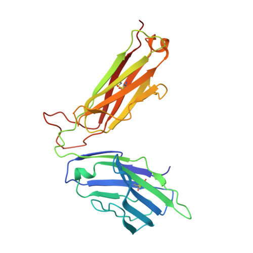
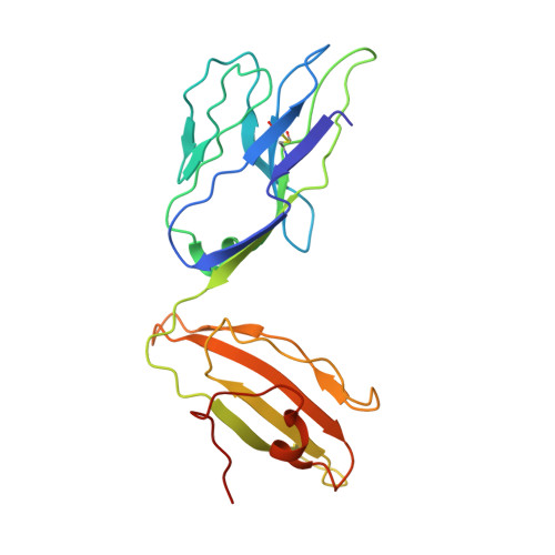
Molecular basis of mycobacterial lipid antigen presentation by CD1c and its recognition by alpha beta T cells.

Roy, S.Ly, D.Li, N.S.Altman, J.D.Piccirilli, J.A.Moody, D.B.Adams, E.J.

(2014) Proc Natl Acad Sci U S A 111: E4648-E4657

  • DOI: https://doi.org/10.1073/pnas.1408549111
  • Primary Citation Related Structures: 
    4ONH, 4ONO

  • PubMed Abstract: 

    CD1c is a member of the group 1 CD1 family of proteins that are specialized for lipid antigen presentation. Despite high cell surface expression of CD1c on key antigen-presenting cells and the discovery of its mycobacterial lipid antigen presentation capability, the molecular basis of CD1c recognition by T cells is unknown. Here we present a comprehensive functional and molecular analysis of αβ T-cell receptor (TCR) recognition of CD1c presenting mycobacterial phosphomycoketide antigens. Our structure of CD1c with the mycobacterial phosphomycoketide (PM) shows similarities to that of CD1c-mannosyl-β1-phosphomycoketide in that the A' pocket accommodates the mycoketide alkyl chain; however, the phosphate head-group of PM is shifted ∼6 Å in relation to that of mannosyl-β1-PM. We also demonstrate a bona fide interaction between six human TCRs and CD1c-mycoketide complexes, measuring high to moderate affinities. The crystal structure of the DN6 TCR and mutagenic studies reveal a requirement of five complementarity determining region (CDR) loops for CD1c recognition. Furthermore, mutagenesis of CD1c reveals residues in both the α1 and α2 helices involved in TCR recognition, yet not entirely overlapping among the examined TCRs. Unlike patterns for MHC I, no archetypical binding footprint is predicted to be shared by CD1c-reactive TCRs, even when recognizing the same or similar antigens.


  • Organizational Affiliation
    • Departments of Biochemistry and Molecular Biology and.

Macromolecule Content 

  • Total Structure Weight: 50.86 kDa 
  • Atom Count: 3,217 
  • Modeled Residue Count: 434 
  • Deposited Residue Count: 448 
  • Unique protein chains: 2

Macromolecules

Find similar proteins by:|  3D Structure
Entity ID: 1
MoleculeChains  Sequence LengthOrganismDetailsImage
T-cell receptor alphaA [auth B]243Homo sapiensMutation(s): 0 
Gene Names: TRAVTRACTRAB
UniProt & NIH Common Fund Data Resources
Find proteins for A0A5B9 (Homo sapiens)
Explore A0A5B9 
Go to UniProtKB:  A0A5B9
PHAROS:  A0A5B9
Entity Groups
Sequence Clusters30% Identity50% Identity70% Identity90% Identity95% Identity100% Identity
UniProt GroupA0A5B9
Glycosylation
Glycosylation Sites: 1Go to GlyGen: A0A5B9-1
Sequence Annotations
Expand
Reference Sequence
Find similar proteins by:|  3D Structure
Entity ID: 2
MoleculeChains  Sequence LengthOrganismDetailsImage
T-cell receptor betaB [auth A]205Homo sapiensMutation(s): 0 
UniProt & NIH Common Fund Data Resources
Find proteins for P01848 (Homo sapiens)
Explore P01848 
Go to UniProtKB:  P01848
PHAROS:  P01848
Entity Groups
Sequence Clusters30% Identity50% Identity70% Identity90% Identity95% Identity100% Identity
UniProt GroupP01848
Glycosylation
Glycosylation Sites: 1Go to GlyGen: P01848-1
Sequence Annotations
Expand
Reference Sequence

Small Molecules

Ligands 3 Unique
IDChains Name / Formula / InChI Key2D Diagram3D Interactions
NAG

Query on NAG


Download:Ideal Coordinates CCD File
C [auth B],
H [auth A]
2-acetamido-2-deoxy-beta-D-glucopyranose
C8 H15 N O6
OVRNDRQMDRJTHS-FMDGEEDCSA-N
GOL

Query on GOL


Download:Ideal Coordinates CCD File
G [auth B],
I [auth A]
GLYCEROL
C3 H8 O3
PEDCQBHIVMGVHV-UHFFFAOYSA-N
CL

Query on CL


Download:Ideal Coordinates CCD File
D [auth B],
E [auth B],
F [auth B]
CHLORIDE ION
Cl
VEXZGXHMUGYJMC-UHFFFAOYSA-M

Experimental Data & Validation

Experimental Data

  • Method: X-RAY DIFFRACTION
  • Resolution: 3.01 Å
  • R-Value Free:  0.317 (Depositor), 0.331 (DCC) 
  • R-Value Work:  0.257 (Depositor), 0.272 (DCC) 
  • R-Value Observed: 0.260 (Depositor) 
Space Group: C 1 2 1
Unit Cell:
Length ( Å )Angle ( ˚ )
a = 143.825α = 90
b = 64.063β = 115.88
c = 69.534γ = 90
Software Package:
Software NamePurpose
DENZOdata reduction
SCALEPACKdata scaling
PHASERphasing
PHENIXrefinement
PDB_EXTRACTdata extraction
HKL-2000data collection

Structure Validation

View Full Validation Report


Ligand Structure Quality Assessment 


Entry History 

Deposition Data

Revision History  (Full details and data files)

  • Version 1.0: 2014-10-08
    Type: Initial release
  • Version 1.1: 2015-02-18
    Changes: Database references
  • Version 1.2: 2020-07-29
    Type: Remediation
    Reason: Carbohydrate remediation
    Changes: Advisory, Data collection, Derived calculations, Structure summary
  • Version 1.3: 2024-11-20
    Changes: Advisory, Data collection, Database references, Structure summary