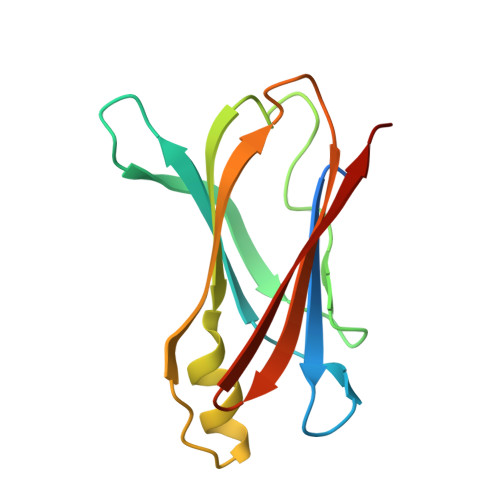
4PVM

Neutron structure of human transthyretin (TTR) at room temperature to 2.0A resolution (Laue)


Experimental Data Snapshot

  • Method: NEUTRON DIFFRACTION
  • Resolution: 2.00 Å
  • R-Value Free: 0.271 
  • R-Value Work: 0.209 
  • R-Value Observed: 0.215 

  • Method: X-RAY DIFFRACTION
  • Resolution: 1.95 Å
  • R-Value Free: 0.203 
  • R-Value Work: 0.153 
  • R-Value Observed: 0.158 

wwPDB Validation   3D Report Full Report


This is version 2.0 of the entry. See complete history


Literature

Binding site asymmetry in human transthyretin: insights from a joint neutron and X-ray crystallographic analysis using perdeuterated protein

Haupt, M.Blakeley, M.P.Fisher, S.J.Mason, S.A.Cooper, J.B.Mitchell, E.P.Forsyth, V.T.

(2014) IUCrJ 1: 429-438

  • DOI: https://doi.org/10.1107/S2052252514021113
  • Primary Citation of Related Structures:  
    4PVL, 4PVM, 4PVN

  • PubMed Abstract: 

    Human transthyretin has an intrinsic tendency to form amyloid fibrils and is heavily implicated in senile systemic amyloidosis. Here, detailed neutron structural studies of perdeuterated transthyretin are described. The analyses, which fully exploit the enhanced visibility of isotopically replaced hydrogen atoms, yield new information on the stability of the protein and the possible mechanisms of amyloid formation. Residue Ser117 may play a pivotal role in that a single water molecule is closely associated with the γ-hydrogen atoms in one of the binding pockets, and could be important in determining which of the two sites is available to the substrate. The hydrogen-bond network at the monomer-monomer interface is more extensive than that at the dimer-dimer interface. Additionally, the edge strands of the primary dimer are seen to be favourable for continuation of the β-sheet and the formation of an extended cross-β structure through sequential dimer couplings. It is argued that the precursor to fibril formation is the dimeric form of the protein.


  • Organizational Affiliation

    Facility of Natural Sciences, Institute of Science and Technology in Medicine, Keele University , Staffordshire ST5 5BG, United Kingdom ; Institut Laue-Langevin , 71, avenue des Martyrs, Grenoble, CS 20156, France ; Partnership for Structural Biology , 71, avenue des Martyrs, Grenoble, CS 20156, France.


Macromolecules
Find similar proteins by:  (by identity cutoff)  |  3D Structure
Entity ID: 1
MoleculeChains Sequence LengthOrganismDetailsImage
Transthyretin
A, B
130Homo sapiensMutation(s): 0 
Gene Names: PALBTTR
UniProt & NIH Common Fund Data Resources
Find proteins for P02766 (Homo sapiens)
Explore P02766 
Go to UniProtKB:  P02766
PHAROS:  P02766
GTEx:  ENSG00000118271 
Entity Groups  
Sequence Clusters30% Identity50% Identity70% Identity90% Identity95% Identity100% Identity
UniProt GroupP02766
Sequence Annotations
Expand
  • Reference Sequence
Experimental Data & Validation

Experimental Data

  • Method: NEUTRON DIFFRACTION
  • Resolution: 2.00 Å
  • R-Value Free: 0.271 
  • R-Value Work: 0.209 
  • R-Value Observed: 0.215 
  • Space Group: P 21 21 2
  • Method: X-RAY DIFFRACTION
  • Resolution: 1.95 Å
  • R-Value Free: 0.203 
  • R-Value Work: 0.153 
  • R-Value Observed: 0.158 
  • Space Group: P 21 21 2
Unit Cell:
Length ( Å )Angle ( ˚ )
a = 43.68α = 90
b = 86.26β = 90
c = 65.72γ = 90
Software Package:
Software NamePurpose
QLDdata collection
PHENIXmodel building
PHENIXrefinement
LAUEGENdata reduction
LSCALEdata scaling
SCALAdata scaling
PHENIXphasing

Structure Validation

View Full Validation Report


Entry History 

Deposition Data

Revision History  (Full details and data files)

  • Version 1.0: 2014-11-12
    Type: Initial release
  • Version 1.1: 2014-12-17
    Changes: Database references
  • Version 1.2: 2017-11-22
    Changes: Data collection, Refinement description
  • Version 2.0: 2024-03-20
    Changes: Atomic model, Data collection, Database references