4QP4 | pdb_00004qp4

Crystal Structure of ERK2 in complex with N-cyclohexyl-9H-purin-6-amine


Experimental Data Snapshot

  • Method: X-RAY DIFFRACTION
  • Resolution: 2.20 Å
  • R-Value Free: 
    0.235 (Depositor), 0.237 (DCC) 
  • R-Value Work: 
    0.187 (Depositor), 0.189 (DCC) 
  • R-Value Observed: 
    0.189 (Depositor) 

wwPDB Validation 3D Report Full Report

Validation slider image for 4QP4

Ligand Structure Quality Assessment 


This is version 1.2 of the entry. See complete history

Literature

Fragment-based discovery of potent ERK2 pyrrolopyrazine inhibitors.

Burdick, D.J.Wang, S.Heise, C.Pan, B.Drummond, J.Yin, J.Goeser, L.Magnuson, S.Blaney, J.Moffat, J.Wang, W.Chen, H.

(2015) Bioorg Med Chem Lett 25: 4728-4732

  • DOI: https://doi.org/10.1016/j.bmcl.2015.08.048
  • Primary Citation Related Structures: 
    4QP1, 4QP2, 4QP3, 4QP4, 4QP6, 4QP7, 4QP8, 4QP9, 4QPA

  • PubMed Abstract: 

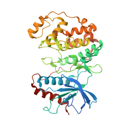
    A fragment-based lead discovery approach was used to discover novel ERK2 inhibitors. The crystal structure of N-benzyl-9H-purin-6-amine 1 in complex with ERK2 elucidated its hinge-binding mode. In addition, the simultaneous binding of an imidazole molecule adjacent to 1 suggested a direction for fragment expansion. Structure-based core hopping applied to 1 led to 5H-pyrrolo[3,2-b]pyrazine (3) that afforded direct vectors to probe the pockets of interest while retaining the essential hinge binding elements. Utilizing the new vectors for SAR exploration, the new core 3 was quickly optimized to compound 39 resulting in a greater than 6600-fold improvement in potency.


  • Organizational Affiliation
    • Department of Discovery Chemistry, Genentech, Inc., South San Francisco, CA 94080, United States. Electronic address: burdick.dan@gene.com.

Macromolecule Content 

  • Total Structure Weight: 85.53 kDa 
  • Atom Count: 5,861 
  • Modeled Residue Count: 672 
  • Deposited Residue Count: 738 
  • Unique protein chains: 1

Macromolecules

Find similar proteins by:|  3D Structure
Entity ID: 1
MoleculeChains  Sequence LengthOrganismDetailsImage
Mitogen-activated protein kinase 1
A, B
369Homo sapiensMutation(s): 0 
Gene Names: MAPK1ERK2PRKM1PRKM2
EC: 2.7.11.24
UniProt & NIH Common Fund Data Resources
Find proteins for P28482 (Homo sapiens)
Explore P28482 
Go to UniProtKB:  P28482
PHAROS:  P28482
GTEx:  ENSG00000100030 
Entity Groups
Sequence Clusters30% Identity50% Identity70% Identity90% Identity95% Identity100% Identity
UniProt GroupP28482
Sequence Annotations
Expand
Reference Sequence

Experimental Data & Validation

Experimental Data

  • Method: X-RAY DIFFRACTION
  • Resolution: 2.20 Å
  • R-Value Free:  0.235 (Depositor), 0.237 (DCC) 
  • R-Value Work:  0.187 (Depositor), 0.189 (DCC) 
  • R-Value Observed: 0.189 (Depositor) 
Space Group: P 1 21 1
Unit Cell:
Length ( Å )Angle ( ˚ )
a = 59.428α = 90
b = 67.229β = 102.21
c = 86.997γ = 90
Software Package:
Software NamePurpose
HKL-2000data collection
PHASERphasing
PHENIXrefinement
DENZOdata reduction
SCALEPACKdata scaling

Structure Validation

View Full Validation Report


Ligand Structure Quality Assessment 


Entry History 

Deposition Data

Revision History  (Full details and data files)

  • Version 1.0: 2015-09-23
    Type: Initial release
  • Version 1.1: 2015-12-16
    Changes: Database references
  • Version 1.2: 2024-02-28
    Changes: Data collection, Database references, Derived calculations