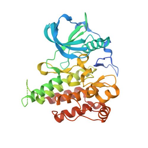
Structure-Guided Blockade of CSF1R Kinase in Tenosynovial Giant-Cell Tumor.
Tap, W.D., Wainberg, Z.A., Anthony, S.P., Ibrahim, P.N., Zhang, C., Healey, J.H., Chmielowski, B., Staddon, A.P., Cohn, A.L., Shapiro, G.I., Keedy, V.L., Singh, A.S., Puzanov, I., Kwak, E.L., Wagner, A.J., Von Hoff, D.D., Weiss, G.J., Ramanathan, R.K., Zhang, J., Habets, G., Zhang, Y., Burton, E.A., Visor, G., Sanftner, L., Severson, P., Nguyen, H., Kim, M.J., Marimuthu, A., Tsang, G., Shellooe, R., Gee, C., West, B.L., Hirth, P., Nolop, K., van de Rijn, M., Hsu, H.H., Peterfy, C., Lin, P.S., Tong-Starksen, S., Bollag, G.(2015) N Engl J Med 373: 428-437
- PubMed: 26222558
- DOI: https://doi.org/10.1056/NEJMoa1411366
- Primary Citation of Related Structures:
4R7H, 4R7I - PubMed Abstract:
Expression of the colony-stimulating factor 1 (CSF1) gene is elevated in most tenosynovial giant-cell tumors. This observation has led to the discovery and clinical development of therapy targeting the CSF1 receptor (CSF1R). Using x-ray co-crystallography to guide our drug-discovery research, we generated a potent, selective CSF1R inhibitor, PLX3397, that traps the kinase in the autoinhibited conformation. We then conducted a multicenter, phase 1 trial in two parts to analyze this compound. In the first part, we evaluated escalations in the dose of PLX3397 that was administered orally in patients with solid tumors (dose-escalation study). In the second part, we evaluated PLX3397 at the chosen phase 2 dose in an extension cohort of patients with tenosynovial giant-cell tumors (extension study). Pharmacokinetic and tumor responses in the enrolled patients were assessed, and CSF1 in situ hybridization was performed to confirm the mechanism of action of PLX3397 and that the pattern of CSF1 expression was consistent with the pathological features of tenosynovial giant-cell tumor. A total of 41 patients were enrolled in the dose-escalation study, and an additional 23 patients were enrolled in the extension study. The chosen phase 2 dose of PLX3397 was 1000 mg per day. In the extension study, 12 patients with tenosynovial giant-cell tumors had a partial response and 7 patients had stable disease. Responses usually occurred within the first 4 months of treatment, and the median duration of response exceeded 8 months. The most common adverse events included fatigue, change in hair color, nausea, dysgeusia, and periorbital edema; adverse events rarely led to discontinuation of treatment. Treatment of tenosynovial giant-cell tumors with PLX3397 resulted in a prolonged regression in tumor volume in most patients. (Funded by Plexxikon; ClinicalTrials.gov number, NCT01004861.).
Organizational Affiliation:
From Memorial Sloan Kettering Cancer Center (W.D.T., J.H.H.) and Weill Cornell Medical College (W.D.T.) - both in New York; University of California, Los Angeles, Medical Center, Los Angeles (Z.A.W., B.C., A.S.S.), Plexxikon, Berkeley (P.N.I., C.Z., J.Z., G.H., Y.Z., E.A.B., G.V., L.S., P.S., H.N., M.J.K., A.M., G.T., R.S., C.G., B.L.W., P.H., K.N., H.H.H., P.S.L., S.T.-S., G.B.), and Stanford University School of Medicine, Stanford (M.R.) - all in California; Evergreen Hematology and Oncology, Spokane, WA (S.P.A.); University of Pennsylvania School of Medicine, Philadelphia (A.P.S.); Rocky Mountain Cancer Centers, Denver (A.L.C.); Dana-Farber Cancer Institute (G.I.S., A.J.W.) and Massachusetts General Hospital (E.L.K.) - both in Boston; Vanderbilt University Medical Center, Nashville (V.L.K., I.P.); Virginia G. Piper Cancer Center at Scottsdale Healthcare-Translational Genomics Research Institute (TGen), Scottsdale, AZ (D.D.V.H., G.J.W., R.K.R.); and Spire Sciences, Boca Raton, FL (C.P.).


