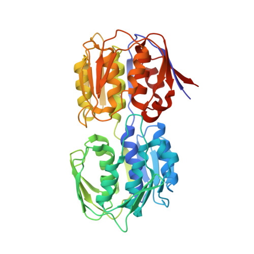
Structure of UDP-N-acetylglucosamine 1-carboxyvinyltransferase from Vibrio cholerae in complex with substrate UDP-N-acetylglucosamine and the drug fosfomycin
Nocek, B., Maltseva, N., Anderson, W., Joachimiak, A., Center for Structural Genomics of Infectious Diseases (CSGID)To be published.