4RL7 | pdb_00004rl7

Crystal structure of Human galectin-3 CRD in complex with lactose (pH 7.5, PEG6000)


Experimental Data Snapshot

  • Method: X-RAY DIFFRACTION
  • Resolution: 2.00 Å
  • R-Value Free: 
    0.245 (Depositor), 0.207 (DCC) 
  • R-Value Work: 
    0.166 (Depositor), 0.185 (DCC) 
  • R-Value Observed: 
    0.173 (Depositor) 

Starting Model: experimental
View more details

wwPDB Validation 3D Report Full Report

Validation slider image for 4RL7

This is version 2.2 of the entry. See complete history

Literature

The water network in galectin-3 ligand binding site guides inhibitor design.

Su, J.Zhang, T.Wang, P.Liu, F.Tai, G.Zhou, Y.

(2015) Acta Biochim Biophys Sin (Shanghai) 47: 192-198

  • DOI: https://doi.org/10.1093/abbs/gmu132
  • Primary Citation Related Structures: 
    4R9A, 4R9B, 4R9C, 4R9D, 4RL7

  • PubMed Abstract: 

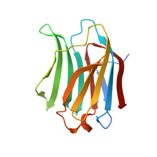
    Galectin-3 (Gal-3) which shows affinity of β-galactosides is a cancer-related protein. Thus, it is important to understand its ligand binding mechanism and then design its specific inhibitor. It was suggested that the positions of water molecules in Gal-3 ligand-binding site could be replaced by appropriate chemical groups of ideal inhibitors. However, the reported structures of Gal-3 carbohydrate recognition domain (CRD) complexed with lactose showed that the number of water molecules are different and the water positions are inconsistent in the ligand-binding site. This study reported four high-resolution (1.24-1.19 Å) structures of Gal-3 CRD complexed with lactose, and accurately located 12 conserved water molecules in the water network of Gal-3 CRD ligand-binding site by merging these structures. These water molecules either directly stabilize the binding of Gal-3 CRD and lactose, or hold the former water molecules at the right place. In particular, water molecule 4 (W4) which only coordinates with water molecule 5 (W5) and water molecule 6 (W6) plays a key role in stabilizing galactose residue. In addition, by three-dimensional alignment of the positions of all residues, 14 flexible parts of Gal-3 CRD were found to dynamically fluctuate in the crystalline environment.


  • Organizational Affiliation
    • School of Life Sciences, Northeast Normal University, Changchun 130024, China.

Macromolecule Content 

  • Total Structure Weight: 16.58 kDa 
  • Atom Count: 1,233 
  • Modeled Residue Count: 138 
  • Deposited Residue Count: 144 
  • Unique protein chains: 1

Macromolecules

Find similar proteins by:|  3D Structure
Entity ID: 1
MoleculeChains  Sequence LengthOrganismDetailsImage
Galectin-3144Homo sapiensMutation(s): 0 
Gene Names: LGALS3MAC2
UniProt & NIH Common Fund Data Resources
Find proteins for P17931 (Homo sapiens)
Explore P17931 
Go to UniProtKB:  P17931
PHAROS:  P17931
GTEx:  ENSG00000131981 
Entity Groups
Sequence Clusters30% Identity50% Identity70% Identity90% Identity95% Identity100% Identity
UniProt GroupP17931
Sequence Annotations
Expand
Reference Sequence

Oligosaccharides

Help  
Entity ID: 2
MoleculeChains Length2D Diagram GlycosylationD Interactions
beta-D-galactopyranose-(1-4)-beta-D-glucopyranose
B
2N/A
Glycosylation Resources
GlyTouCan: G84224TW
GlyCosmos: G84224TW
GlyGen: G84224TW

Biologically Interesting Molecules (External Reference) 

1 Unique

Experimental Data & Validation

Experimental Data

  • Method: X-RAY DIFFRACTION
  • Resolution: 2.00 Å
  • R-Value Free:  0.245 (Depositor), 0.207 (DCC) 
  • R-Value Work:  0.166 (Depositor), 0.185 (DCC) 
  • R-Value Observed: 0.173 (Depositor) 
Space Group: P 21 21 21
Unit Cell:
Length ( Å )Angle ( ˚ )
a = 36.2α = 90
b = 57.8β = 90
c = 63γ = 90
Software Package:
Software NamePurpose
PHENIXrefinement

Structure Validation

View Full Validation Report


Entry History 

Deposition Data

  • Released Date: 2015-03-11 
  • Deposition Author(s): Su, J.Y.

Revision History  (Full details and data files)

  • Version 1.0: 2015-03-11
    Type: Initial release
  • Version 2.0: 2020-07-29
    Type: Remediation
    Reason: Carbohydrate remediation
    Changes: Atomic model, Data collection, Database references, Derived calculations, Non-polymer description, Structure summary
  • Version 2.1: 2022-08-24
    Changes: Database references, Structure summary
  • Version 2.2: 2023-11-08
    Changes: Data collection, Refinement description