4RSE | pdb_00004rse

Crystal structure of RPE65 in complex with MB-001 and palmitate


Experimental Data Snapshot

  • Method: X-RAY DIFFRACTION
  • Resolution: 2.39 Å
  • R-Value Free: 
    0.236 (Depositor), 0.236 (DCC) 
  • R-Value Work: 
    0.196 (Depositor), 0.201 (DCC) 
  • R-Value Observed: 
    0.198 (Depositor) 

Starting Model: experimental
View more details

wwPDB Validation 3D Report Full Report

Validation slider image for 4RSE

Ligand Structure Quality Assessment 


This is version 1.3 of the entry. See complete history

Literature

Catalytic mechanism of a retinoid isomerase essential for vertebrate vision.

Kiser, P.D.Zhang, J.Badiee, M.Li, Q.Shi, W.Sui, X.Golczak, M.Tochtrop, G.P.Palczewski, K.

(2015) Nat Chem Biol 11: 409-415

  • DOI: https://doi.org/10.1038/nchembio.1799
  • Primary Citation Related Structures: 
    4RSC, 4RSE

  • PubMed Abstract: 

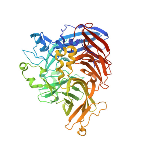
    Visual function in vertebrates is dependent on the membrane-bound retinoid isomerase RPE65, an essential component of the retinoid cycle pathway that regenerates 11-cis-retinal for rod and cone opsins. The mechanism by which RPE65 catalyzes stereoselective retinoid isomerization has remained elusive because of uncertainty about how retinoids bind to its active site. Here we present crystal structures of RPE65 in complex with retinoid-mimetic compounds, one of which is in clinical trials for the treatment of age-related macular degeneration. The structures reveal the active site retinoid-binding cavity located near the membrane-interacting surface of the enzyme as well as an Fe-bound palmitate ligand positioned in an adjacent pocket. With the geometry of the RPE65-substrate complex clarified, we delineate a mechanism of catalysis that reconciles the extensive biochemical and structural research on this enzyme. These data provide molecular foundations for understanding a key process in vision and pharmacological inhibition of RPE65 with small molecules.


  • Organizational Affiliation
    • Department of Pharmacology, Cleveland Center for Membrane and Structural Biology, School of Medicine, Case Western Reserve University, Cleveland, Ohio, USA.

Macromolecule Content 

  • Total Structure Weight: 123.1 kDa 
  • Atom Count: 8,780 
  • Modeled Residue Count: 1,022 
  • Deposited Residue Count: 1,066 
  • Unique protein chains: 1

Macromolecules

Find similar proteins by:|  3D Structure
Entity ID: 1
MoleculeChains  Sequence LengthOrganismDetailsImage
Retinoid isomerohydrolase
A, B
533Bos taurusMutation(s): 0 
EC: 3.1.1.64 (PDB Primary Data), 5.3.3.22 (UniProt)
UniProt
Find proteins for Q28175 (Bos taurus)
Explore Q28175 
Go to UniProtKB:  Q28175
Entity Groups
Sequence Clusters30% Identity50% Identity70% Identity90% Identity95% Identity100% Identity
UniProt GroupQ28175
Sequence Annotations
Expand
Reference Sequence

Small Molecules

Binding Affinity Annotations 
IDSourceBinding Affinity
A6V BindingDB:  4RSE IC50: min: 323, max: 323 (nM) from 2 assay(s)
EC50: 323 (nM) from 1 assay(s)

Experimental Data & Validation

Experimental Data

  • Method: X-RAY DIFFRACTION
  • Resolution: 2.39 Å
  • R-Value Free:  0.236 (Depositor), 0.236 (DCC) 
  • R-Value Work:  0.196 (Depositor), 0.201 (DCC) 
  • R-Value Observed: 0.198 (Depositor) 
Space Group: P 65
Unit Cell:
Length ( Å )Angle ( ˚ )
a = 175.639α = 90
b = 175.639β = 90
c = 86.611γ = 120
Software Package:
Software NamePurpose
XDSdata scaling
REFMACrefinement
XDSdata reduction
REFMACphasing

Structure Validation

View Full Validation Report


Ligand Structure Quality Assessment 


Entry History 

Deposition Data

Revision History  (Full details and data files)

  • Version 1.0: 2015-04-15
    Type: Initial release
  • Version 1.1: 2015-06-03
    Changes: Database references
  • Version 1.2: 2023-09-20
    Changes: Data collection, Database references, Derived calculations, Refinement description
  • Version 1.3: 2024-11-06
    Changes: Structure summary