4V3W | pdb_00004v3w

Structure of rat neuronal nitric oxide synthase heme domain in complex with 2-(2-(1H-imidazol-1-yl)pyrimidin-4-yl)-N-(3- fluorophenethyl)ethan-1-amine


Experimental Data Snapshot

  • Method: X-RAY DIFFRACTION
  • Resolution: 2.13 Å
  • R-Value Free: 
    0.265 (Depositor), 0.264 (DCC) 
  • R-Value Work: 
    0.206 (Depositor), 0.206 (DCC) 
  • R-Value Observed: 
    0.209 (Depositor) 

wwPDB Validation 3D Report Full Report

Validation slider image for 4V3W

Ligand Structure Quality Assessment 


This is version 1.2 of the entry. See complete history

Literature

Novel 2,4-Disubstituted Pyrimidines as Potent, Selective, and Cell-Permeable Inhibitors of Neuronal Nitric Oxide Synthase.

Mukherjee, P.Li, H.Sevrioukova, I.Chreifi, G.Martasek, P.Roman, L.J.Poulos, T.L.Silverman, R.B.

(2015) J Med Chem 58: 1067

  • DOI: https://doi.org/10.1021/jm501719e
  • Primary Citation Related Structures: 
    4D2Y, 4D2Z, 4D30, 4D31, 4D32, 4D33, 4D34, 4D35, 4D36, 4D37, 4D38, 4D39, 4D3A, 4D3B, 4UCH, 4V3U, 4V3V, 4V3W, 4V3X, 4V3Y, 4V3Z

  • PubMed Abstract: 

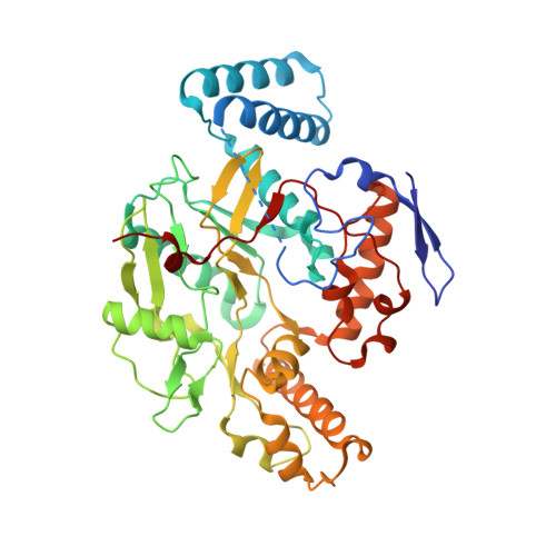
    Selective inhibition of neuronal nitric oxide synthase (nNOS) is an important therapeutic approach to target neurodegenerative disorders. However, the majority of the nNOS inhibitors developed are arginine mimetics and, therefore, suffer from poor bioavailability. We designed a novel strategy to combine a more pharmacokinetically favorable 2-imidazolylpyrimidine head with promising structural components from previous inhibitors. In conjunction with extensive structure-activity studies, several highly potent and selective inhibitors of nNOS were discovered. X-ray crystallographic analysis reveals that these type II inhibitors utilize the same hydrophobic pocket to gain strong inhibitory potency (13), as well as high isoform selectivity. Interestingly, select compounds from this series (9) showed good permeability and low efflux in a Caco-2 assay, suggesting potential oral bioavailability, and exhibited minimal off-target binding to 50 central nervous system receptors. Furthermore, even with heme-coordinating groups in the molecule, modifying other pharmacophoric fragments minimized undesirable inhibition of cytochrome P450s from human liver microsomes.


  • Organizational Affiliation
    • Department of Chemistry, Department of Molecular Biosciences, Chemistry of Life Processes Institute, Center for Molecular Innovation and Drug Discovery, Northwestern University , Evanston, Illinois 60208-3113, United States.

Macromolecule Content 

  • Total Structure Weight: 100.15 kDa 
  • Atom Count: 7,004 
  • Modeled Residue Count: 819 
  • Deposited Residue Count: 844 
  • Unique protein chains: 1

Macromolecules

Find similar proteins by:|  3D Structure
Entity ID: 1
MoleculeChains  Sequence LengthOrganismDetailsImage
NITRIC OXIDE SYNTHASE, BRAIN
A, B
422Rattus norvegicusMutation(s): 0 
EC: 1.14.13.39
UniProt
Find proteins for P29476 (Rattus norvegicus)
Explore P29476 
Go to UniProtKB:  P29476
Entity Groups
Sequence Clusters30% Identity50% Identity70% Identity90% Identity95% Identity100% Identity
UniProt GroupP29476
Sequence Annotations
Expand
Reference Sequence

Small Molecules

Ligands 5 Unique
IDChains Name / Formula / InChI Key2D Diagram3D Interactions
HEM

Query on HEM


Download:Ideal Coordinates CCD File
C [auth A],
H [auth B]
PROTOPORPHYRIN IX CONTAINING FE
C34 H32 Fe N4 O4
KABFMIBPWCXCRK-RGGAHWMASA-L
E2Z

Query on E2Z


Download:Ideal Coordinates CCD File
E [auth A],
J [auth B]
2-(3-fluorophenyl)-N-{2-[2-(1H-imidazol-1-yl)pyrimidin-4-yl]ethyl}ethanamine
C17 H18 F N5
ZPTNPJCYGBTULC-UHFFFAOYSA-N
H4B

Query on H4B


Download:Ideal Coordinates CCD File
D [auth A],
I [auth B]
5,6,7,8-TETRAHYDROBIOPTERIN
C9 H15 N5 O3
FNKQXYHWGSIFBK-RPDRRWSUSA-N
ZN

Query on ZN


Download:Ideal Coordinates CCD File
G [auth A]ZINC ION
Zn
PTFCDOFLOPIGGS-UHFFFAOYSA-N
ACT

Query on ACT


Download:Ideal Coordinates CCD File
F [auth A],
K [auth B]
ACETATE ION
C2 H3 O2
QTBSBXVTEAMEQO-UHFFFAOYSA-M

Experimental Data & Validation

Experimental Data

  • Method: X-RAY DIFFRACTION
  • Resolution: 2.13 Å
  • R-Value Free:  0.265 (Depositor), 0.264 (DCC) 
  • R-Value Work:  0.206 (Depositor), 0.206 (DCC) 
  • R-Value Observed: 0.209 (Depositor) 
Space Group: P 21 21 21
Unit Cell:
Length ( Å )Angle ( ˚ )
a = 51.837α = 90
b = 110.25β = 90
c = 164.318γ = 90
Software Package:
Software NamePurpose
REFMACrefinement
XDSdata reduction
SCALAdata scaling
REFMACphasing

Structure Validation

View Full Validation Report


Ligand Structure Quality Assessment 


Entry History 

Deposition Data

Revision History  (Full details and data files)

  • Version 1.0: 2014-12-24
    Type: Initial release
  • Version 1.1: 2015-03-04
    Changes: Database references
  • Version 1.2: 2024-05-08
    Changes: Data collection, Database references, Derived calculations, Other