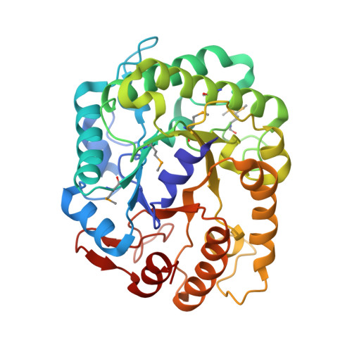
4W88

Crystal structure of XEG5A, a GH5 xyloglucan-specific endo-beta-1,4-glucanase from ruminal metagenomic library, in complex with a xyloglucan oligosaccharide and TRIS


Experimental Data Snapshot

  • Method: X-RAY DIFFRACTION
  • Resolution: 1.58 Å
  • R-Value Free: 0.162 
  • R-Value Work: 0.125 
  • R-Value Observed: 0.127 

wwPDB Validation   3D Report Full Report


This is version 2.1 of the entry. See complete history


Literature

Structural Basis for Xyloglucan Specificity and alpha-d-Xylp(1 6)-d-Glcp Recognition at the -1 Subsite within the GH5 Family.

Dos Santos, C.R.Cordeiro, R.L.Wong, D.W.Murakami, M.T.

(2015) Biochemistry 54: 1930-1942

  • DOI: https://doi.org/10.1021/acs.biochem.5b00011
  • Primary Citation of Related Structures:  
    4W84, 4W85, 4W86, 4W87, 4W88, 4W89, 4W8A, 4W8B

  • PubMed Abstract: 

    GH5 is one of the largest glycoside hydrolase families, comprising at least 20 distinct activities within a common structural scaffold. However, the molecular basis for the functional differentiation among GH5 members is still not fully understood, principally for xyloglucan specificity. In this work, we elucidated the crystal structures of two novel GH5 xyloglucanases (XEGs) retrieved from a rumen microflora metagenomic library, in the native state and in complex with xyloglucan-derived oligosaccharides. These results provided insights into the structural determinants that differentiate GH5 XEGs from parental cellulases and a new mode of action within the GH5 family related to structural adaptations in the -1 subsite. The oligosaccharide found in the XEG5A complex, permitted the mapping, for the first time, of the positive subsites of a GH5 XEG, revealing the importance of the pocket-like topology of the +1 subsite in conferring the ability of some GH5 enzymes to attack xyloglucan. Complementarily, the XEG5B complex covered the negative subsites, completing the subsite mapping of GH5 XEGs at high resolution. Interestingly, XEG5B is, to date, the only GH5 member able to cleave XXXG into XX and XG, and in the light of these results, we propose that a modification in the -1 subsite enables the accommodation of a xylosyl side chain at this position. The stereochemical compatibility of the -1 subsite with a xylosyl moiety was also reported for other structurally nonrelated XEGs belonging to the GH74 family, indicating it to be an essential attribute for this mode of action.


  • Organizational Affiliation

    †Brazilian Biosciences National Laboratory, National Center of Research in Energy and Materials, Campinas, São Paulo 13083-970, Brazil.


Macromolecules
Find similar proteins by:  (by identity cutoff)  |  3D Structure
Entity ID: 1
MoleculeChains Sequence LengthOrganismDetailsImage
Xyloglucan-specific endo-beta-1,4-glucanaseA [auth B],
B [auth A]
340uncultured bacteriumMutation(s): 1 
EC: 3.2.1.151
UniProt
Find proteins for D2K7Z0 (uncultured bacterium)
Explore D2K7Z0 
Go to UniProtKB:  D2K7Z0
Entity Groups  
Sequence Clusters30% Identity50% Identity70% Identity90% Identity95% Identity100% Identity
UniProt GroupD2K7Z0
Sequence Annotations
Expand
  • Reference Sequence
Oligosaccharides

Help

Entity ID: 2
MoleculeChains Length2D Diagram Glycosylation3D Interactions
beta-D-galactopyranose-(1-2)-alpha-D-xylopyranose-(1-6)-[beta-D-glucopyranose-(1-4)]beta-D-glucopyranose
C
4N/A
Glycosylation Resources
GlyTouCan:  G23047PZ
GlyCosmos:  G23047PZ
Entity ID: 3
MoleculeChains Length2D Diagram Glycosylation3D Interactions
alpha-D-xylopyranose-(1-6)-beta-D-glucopyranose-(1-4)-[beta-D-galactopyranose-(1-2)-alpha-D-xylopyranose-(1-6)]beta-D-glucopyranose-(1-4)-beta-D-glucopyranose
D
6N/A
Glycosylation Resources
GlyTouCan:  G23873IA
GlyCosmos:  G23873IA
Small Molecules
Modified Residues  1 Unique
IDChains TypeFormula2D DiagramParent
MSE
Query on MSE
A [auth B],
B [auth A]
L-PEPTIDE LINKINGC5 H11 N O2 SeMET
Experimental Data & Validation

Experimental Data

  • Method: X-RAY DIFFRACTION
  • Resolution: 1.58 Å
  • R-Value Free: 0.162 
  • R-Value Work: 0.125 
  • R-Value Observed: 0.127 
  • Space Group: P 31
Unit Cell:
Length ( Å )Angle ( ˚ )
a = 97.658α = 90
b = 97.658β = 90
c = 95.985γ = 120
Software Package:
Software NamePurpose
REFMACrefinement
XDSdata scaling
MOLREPphasing

Structure Validation

View Full Validation Report


Entry History & Funding Information

Deposition Data


Funding OrganizationLocationGrant Number
Sao Paulo Research Foundation (FAPESP)Brazil2013/13309-0; 201/07135-1

Revision History  (Full details and data files)

  • Version 1.0: 2015-03-11
    Type: Initial release
  • Version 1.1: 2015-03-25
    Changes: Database references
  • Version 1.2: 2017-11-22
    Changes: Database references, Derived calculations, Refinement description, Source and taxonomy
  • Version 1.3: 2019-04-17
    Changes: Author supporting evidence, Data collection
  • Version 1.4: 2020-01-01
    Changes: Author supporting evidence, Data collection
  • Version 2.0: 2020-07-29
    Type: Remediation
    Reason: Carbohydrate remediation
    Changes: Advisory, Atomic model, Data collection, Derived calculations, Structure summary
  • Version 2.1: 2023-12-27
    Changes: Data collection, Database references, Derived calculations, Structure summary