4Y6G

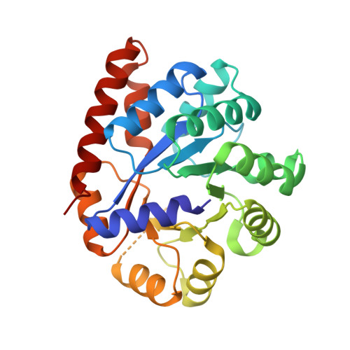
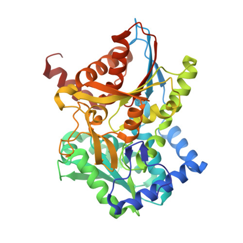
Crystal structure of Tryptophan Synthase from Salmonella typhimurium in complex with N-(4'-trifluoromethoxybenzoyl)-2-amino-1-ethylphosphate (F6F) inhibitor in the alpha-site and beta-site.


Experimental Data Snapshot

  • Method: X-RAY DIFFRACTION
  • Resolution: 1.65 Å
  • R-Value Free: 0.179 
  • R-Value Work: 0.158 
  • R-Value Observed: 0.159 

Starting Model: experimental
View more details

wwPDB Validation   3D Report Full Report


Ligand Structure Quality Assessment 


This is version 1.3 of the entry. See complete history


Literature

Visualizing the tunnel in tryptophan synthase with crystallography: Insights into a selective filter for accommodating indole and rejecting water.

Hilario, E.Caulkins, B.G.Huang, Y.M.You, W.Chang, C.E.Mueller, L.J.Dunn, M.F.Fan, L.

(2016) Biochim Biophys Acta 1864: 268-279

  • DOI: https://doi.org/10.1016/j.bbapap.2015.12.006
  • Primary Citation of Related Structures:  
    4WX2, 4Y6G, 4ZQC, 5BW6

  • PubMed Abstract: 

    Four new X-ray structures of tryptophan synthase (TS) crystallized with varying numbers of the amphipathic N-(4'-trifluoromethoxybenzoyl)-2-aminoethyl phosphate (F6) molecule are presented. These structures show one of the F6 ligands threaded into the tunnel from the β-site and reveal a distinct hydrophobic region. Over this expanse, the interactions between F6 and the tunnel are primarily nonpolar, while the F6 phosphoryl group fits into a polar pocket of the β-subunit active site. Further examination of TS structures reveals that one portion of the tunnel (T1) binds clusters of water molecules, whereas waters are not observed in the nonpolar F6 binding region of the tunnel (T2). MD simulation of another TS structure with an unobstructed tunnel also indicates the T2 region of the tunnel excludes water, consistent with a dewetted state that presents a significant barrier to the transfer of water into the closed β-site. We conclude that hydrophobic molecules can freely diffuse between the α- and β-sites via the tunnel, while water does not. We propose that exclusion of water serves to inhibit reaction of water with the α-aminoacrylate intermediate to form ammonium ion and pyruvate, a deleterious side reaction in the αβ-catalytic cycle. Finally, while most TS structures show βPhe280 partially blocking the tunnel between the α- and β-sites, new structures show an open tunnel, suggesting the flexibility of the βPhe280 side chain. Flexible docking studies and MD simulations confirm that the dynamic behavior of βPhe280 allows unhindered transfer of indole through the tunnel, therefore excluding a gating role for this residue.


  • Organizational Affiliation

    Department of Biochemistry, University of California at Riverside, Riverside, CA 92521, USA.


Macromolecules
Find similar proteins by:  (by identity cutoff)  |  3D Structure
Entity ID: 1
MoleculeChains Sequence LengthOrganismDetailsImage
Tryptophan synthase alpha chain268Salmonella enterica subsp. enterica serovar TyphimuriumMutation(s): 0 
Gene Names: trpA
EC: 4.2.1.20
UniProt
Find proteins for P00929 (Salmonella typhimurium (strain LT2 / SGSC1412 / ATCC 700720))
Explore P00929 
Go to UniProtKB:  P00929
Entity Groups  
Sequence Clusters30% Identity50% Identity70% Identity90% Identity95% Identity100% Identity
UniProt GroupP00929
Sequence Annotations
Expand
  • Reference Sequence
Find similar proteins by:  (by identity cutoff)  |  3D Structure
Entity ID: 2
MoleculeChains Sequence LengthOrganismDetailsImage
Tryptophan synthase beta chain397Salmonella enterica subsp. enterica serovar TyphimuriumMutation(s): 0 
Gene Names: trpB
EC: 4.2.1.20
UniProt
Find proteins for P0A2K1 (Salmonella typhimurium (strain LT2 / SGSC1412 / ATCC 700720))
Explore P0A2K1 
Go to UniProtKB:  P0A2K1
Entity Groups  
Sequence Clusters30% Identity50% Identity70% Identity90% Identity95% Identity100% Identity
UniProt GroupP0A2K1
Sequence Annotations
Expand
  • Reference Sequence
Experimental Data & Validation

Experimental Data

  • Method: X-RAY DIFFRACTION
  • Resolution: 1.65 Å
  • R-Value Free: 0.179 
  • R-Value Work: 0.158 
  • R-Value Observed: 0.159 
  • Space Group: C 1 2 1
Unit Cell:
Length ( Å )Angle ( ˚ )
a = 184.17α = 90
b = 59.84β = 94.91
c = 67.3γ = 90
Software Package:
Software NamePurpose
REFMACrefinement
SCALAdata scaling
MOLREPphasing
PDB_EXTRACTdata extraction
MOSFLMdata reduction

Structure Validation

View Full Validation Report


Ligand Structure Quality Assessment 


Entry History & Funding Information

Deposition Data


Funding OrganizationLocationGrant Number
National Institutes of Health/National Institute of General Medical Sciences (NIH/NIGMS)United StatesR01GM097569

Revision History  (Full details and data files)

  • Version 1.0: 2016-02-10
    Type: Initial release
  • Version 1.1: 2017-09-27
    Changes: Author supporting evidence, Database references, Derived calculations, Refinement description
  • Version 1.2: 2019-12-25
    Changes: Author supporting evidence
  • Version 1.3: 2023-09-27
    Changes: Data collection, Database references, Derived calculations, Refinement description