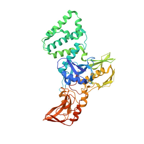
4Z32

Crystal Structure of the FERM-SH2 Domains of Jak2


Experimental Data Snapshot

  • Method: X-RAY DIFFRACTION
  • Resolution: 3.04 Å
  • R-Value Free: 0.276 
  • R-Value Work: 0.257 
  • R-Value Observed: 0.258 

Starting Model: experimental
View more details

wwPDB Validation   3D Report Full Report


This is version 1.1 of the entry. See complete history


Literature

Crystal Structure of the FERM-SH2 Module of Human Jak2.

McNally, R.Toms, A.V.Eck, M.J.

(2016) PLoS One 11: e0156218-e0156218

  • DOI: https://doi.org/10.1371/journal.pone.0156218
  • Primary Citation of Related Structures:  
    4Z32

  • PubMed Abstract: 

    Jak-family tyrosine kinases mediate signaling from diverse cytokine receptors. Binding of Jaks to their cognate receptors is mediated by their N-terminal region, which contains FERM and SH2 domains. Here we describe the crystal structure of the FERM-SH2 region of Jak2 at 3.0Å resolution. The structure reveals that these domains and their flanking linker segments interact intimately to form an integrated structural module. The Jak2 FERM-SH2 structure closely resembles that recently described for Tyk2, another member of the Jak family. While the overall architecture and interdomain orientations are preserved between Jak2 and Tyk2, we identify residues in the putative receptor-binding groove that differ between the two and may contribute to the specificity of receptor recognition. Analysis of Jak mutations that are reported to disrupt receptor binding reveals that they lie in the hydrophobic core of the FERM domain, and are thus expected to compromise the structural integrity of the FERM-SH2 unit. Similarly, analysis of mutations in Jak3 that are associated with severe combined immunodeficiency suggests that they compromise Jak3 function by destabilizing the FERM-SH2 structure.


  • Organizational Affiliation

    Department of Cancer Biology, Dana-Farber Cancer Institute, Boston, Massachusetts, United States of America.


Macromolecules
Find similar proteins by:  (by identity cutoff)  |  3D Structure
Entity ID: 1
MoleculeChains Sequence LengthOrganismDetailsImage
Tyrosine-protein kinase JAK2
A, B, C, D, E
A, B, C, D, E, F, G, H
497Homo sapiensMutation(s): 0 
Gene Names: JAK2
EC: 2.7.10.2
UniProt & NIH Common Fund Data Resources
Find proteins for O60674 (Homo sapiens)
Explore O60674 
Go to UniProtKB:  O60674
PHAROS:  O60674
GTEx:  ENSG00000096968 
Entity Groups  
Sequence Clusters30% Identity50% Identity70% Identity90% Identity95% Identity100% Identity
UniProt GroupO60674
Sequence Annotations
Expand
  • Reference Sequence
Experimental Data & Validation

Experimental Data

  • Method: X-RAY DIFFRACTION
  • Resolution: 3.04 Å
  • R-Value Free: 0.276 
  • R-Value Work: 0.257 
  • R-Value Observed: 0.258 
  • Space Group: P 1 21 1
Unit Cell:
Length ( Å )Angle ( ˚ )
a = 118.193α = 90
b = 188.742β = 113.87
c = 118.585γ = 90
Software Package:
Software NamePurpose
PHENIXrefinement
XDSdata reduction
SCALAdata scaling
PHASERphasing

Structure Validation

View Full Validation Report


Entry History 

Deposition Data

Revision History  (Full details and data files)

  • Version 1.0: 2016-06-22
    Type: Initial release
  • Version 1.1: 2023-09-27
    Changes: Data collection, Database references, Derived calculations, Refinement description