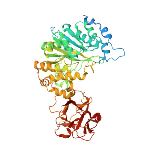
5AJP

Crystal structure of the active form of GalNAc-T2 in complex with UDP and the glycopeptide MUC5AC-13


Experimental Data Snapshot

  • Method: X-RAY DIFFRACTION
  • Resolution: 1.65 Å
  • R-Value Free: 0.186 
  • R-Value Work: 0.156 
  • R-Value Observed: 0.157 

Starting Model: experimental
View more details

wwPDB Validation   3D Report Full Report


Ligand Structure Quality Assessment 


This is version 2.1 of the entry. See complete history


Literature

Dynamic Interplay between Catalytic and Lectin Domains of Galnac-Transferases Modulates Protein O-Glycosylation.

Lira-Navarrete, E.De Las Rivas, M.Companon, I.Pallares, M.C.Kong, Y.Iglesias-Fernandez, J.Bernardes, G.J.L.Peregrina, J.M.Rovira, C.Bernado, P.Bruscolini, P.Clausen, H.Lostao, A.Corzana, F.Hurtado-Guerrero, R.

(2015) Nat Commun 6: 6937

  • DOI: https://doi.org/10.1038/ncomms7937
  • Primary Citation of Related Structures:  
    5AJN, 5AJO, 5AJP

  • PubMed Abstract: 

    Protein O-glycosylation is controlled by polypeptide GalNAc-transferases (GalNAc-Ts) that uniquely feature both a catalytic and lectin domain. The underlying molecular basis of how the lectin domains of GalNAc-Ts contribute to glycopeptide specificity and catalysis remains unclear. Here we present the first crystal structures of complexes of GalNAc-T2 with glycopeptides that together with enhanced sampling molecular dynamics simulations demonstrate a cooperative mechanism by which the lectin domain enables free acceptor sites binding of glycopeptides into the catalytic domain. Atomic force microscopy and small-angle X-ray scattering experiments further reveal a dynamic conformational landscape of GalNAc-T2 and a prominent role of compact structures that are both required for efficient catalysis. Our model indicates that the activity profile of GalNAc-T2 is dictated by conformational heterogeneity and relies on a flexible linker located between the catalytic and the lectin domains. Our results also shed light on how GalNAc-Ts generate dense decoration of proteins with O-glycans.


  • Organizational Affiliation

    BIFI, University of Zaragoza, BIFI-IQFR (CSIC) Joint Unit, Mariano Esquillor s/n, Campus Rio Ebro, Edificio I+D, Zaragoza 50018, Spain.


Macromolecules
Find similar proteins by:  (by identity cutoff)  |  3D Structure
Entity ID: 1
MoleculeChains Sequence LengthOrganismDetailsImage
POLYPEPTIDE N-ACETYLGALACTOSAMINYLTRANSFERASE 2571Homo sapiensMutation(s): 1 
EC: 2.4.1.41
UniProt & NIH Common Fund Data Resources
Find proteins for Q10471 (Homo sapiens)
Explore Q10471 
Go to UniProtKB:  Q10471
PHAROS:  Q10471
GTEx:  ENSG00000143641 
Entity Groups  
Sequence Clusters30% Identity50% Identity70% Identity90% Identity95% Identity100% Identity
UniProt GroupQ10471
Sequence Annotations
Expand
  • Reference Sequence

Find similar proteins by:  Sequence   |   3D Structure  

Entity ID: 2
MoleculeChains Sequence LengthOrganismDetailsImage
MUCIN17Homo sapiensMutation(s): 0 
UniProt & NIH Common Fund Data Resources
Find proteins for P98088 (Homo sapiens)
Explore P98088 
Go to UniProtKB:  P98088
PHAROS:  P98088
GTEx:  ENSG00000215182 
Entity Groups  
Sequence Clusters30% Identity50% Identity70% Identity90% Identity95% Identity100% Identity
UniProt GroupP98088
Glycosylation
Glycosylation Sites: 1Go to GlyGen: P98088-1
Sequence Annotations
Expand
  • Reference Sequence
Small Molecules
Ligands 4 Unique
IDChains Name / Formula / InChI Key2D Diagram3D Interactions
UDP
Query on UDP

Download Ideal Coordinates CCD File 
C [auth A]URIDINE-5'-DIPHOSPHATE
C9 H14 N2 O12 P2
XCCTYIAWTASOJW-XVFCMESISA-N
A2G
Query on A2G

Download Ideal Coordinates CCD File 
Z [auth B]2-acetamido-2-deoxy-alpha-D-galactopyranose
C8 H15 N O6
OVRNDRQMDRJTHS-CBQIKETKSA-N
SO4
Query on SO4

Download Ideal Coordinates CCD File 
D [auth A]
F [auth A]
G [auth A]
H [auth A]
I [auth A]
D [auth A],
F [auth A],
G [auth A],
H [auth A],
I [auth A],
J [auth A],
K [auth A],
L [auth A],
M [auth A],
N [auth A],
O [auth A],
P [auth A],
Q [auth A],
R [auth A],
S [auth A],
T [auth A],
U [auth A],
V [auth A],
W [auth A],
X [auth A],
Y [auth A]
SULFATE ION
O4 S
QAOWNCQODCNURD-UHFFFAOYSA-L
MN
Query on MN

Download Ideal Coordinates CCD File 
E [auth A]MANGANESE (II) ION
Mn
WAEMQWOKJMHJLA-UHFFFAOYSA-N
Experimental Data & Validation

Experimental Data

  • Method: X-RAY DIFFRACTION
  • Resolution: 1.65 Å
  • R-Value Free: 0.186 
  • R-Value Work: 0.156 
  • R-Value Observed: 0.157 
  • Space Group: I 41
Unit Cell:
Length ( Å )Angle ( ˚ )
a = 87.266α = 90
b = 87.266β = 90
c = 178.59γ = 90
Software Package:
Software NamePurpose
REFMACrefinement
XDSdata reduction
SCALEPACKdata scaling
MOLREPphasing

Structure Validation

View Full Validation Report


Ligand Structure Quality Assessment 


Entry History 

Revision History  (Full details and data files)

  • Version 1.0: 2015-03-11
    Type: Initial release
  • Version 1.1: 2015-05-20
    Changes: Database references
  • Version 2.0: 2020-07-29
    Type: Remediation
    Reason: Carbohydrate remediation
    Changes: Atomic model, Data collection, Derived calculations, Other, Structure summary
  • Version 2.1: 2024-01-10
    Changes: Data collection, Database references, Refinement description, Structure summary