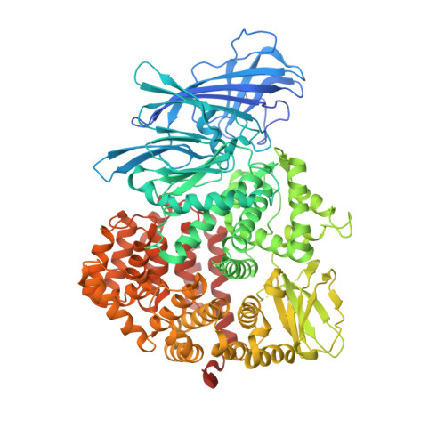
5C97

Insulin regulated aminopeptidase


Experimental Data Snapshot

  • Method: X-RAY DIFFRACTION
  • Resolution: 3.37 Å
  • R-Value Free: 0.285 
  • R-Value Work: 0.216 
  • R-Value Observed: 0.220 

Starting Model: experimental
View more details

wwPDB Validation   3D Report Full Report


Ligand Structure Quality Assessment 


This is version 2.1 of the entry. See complete history


Literature

Crystal Structure of Insulin-Regulated Aminopeptidase with Bound Substrate Analogue Provides Insight on Antigenic Epitope Precursor Recognition and Processing.

Mpakali, A.Saridakis, E.Harlos, K.Zhao, Y.Papakyriakou, A.Kokkala, P.Georgiadis, D.Stratikos, E.

(2015) J Immunol 195: 2842-2851

  • DOI: https://doi.org/10.4049/jimmunol.1501103
  • Primary Citation of Related Structures:  
    4Z7I, 5C97

  • PubMed Abstract: 

    Aminopeptidases that generate antigenic peptides influence immunodominance and adaptive cytotoxic immune responses. The mechanisms that allow these enzymes to efficiently process a vast number of different long peptide substrates are poorly understood. In this work, we report the structure of insulin-regulated aminopeptidase, an enzyme that prepares antigenic epitopes for cross-presentation in dendritic cells, in complex with an antigenic peptide precursor analog. Insulin-regulated aminopeptidase is found in a semiclosed conformation with an extended internal cavity with limited access to the solvent. The N-terminal moiety of the peptide is located at the active site, positioned optimally for catalysis, whereas the C-terminal moiety of the peptide is stabilized along the extended internal cavity lodged between domains II and IV. Hydrophobic interactions and shape complementarity enhance peptide affinity beyond the catalytic site and support a limited selectivity model for antigenic peptide selection that may underlie the generation of complex immunopeptidomes.


  • Organizational Affiliation

    National Center for Scientific Research Demokritos, Agia Paraskevi, Athens 15310, Greece;


Macromolecules
Find similar proteins by:  (by identity cutoff)  |  3D Structure
Entity ID: 1
MoleculeChains Sequence LengthOrganismDetailsImage
Leucyl-cystinyl aminopeptidase
A, B
912Homo sapiensMutation(s): 0 
Gene Names: LNPEPOTASE
EC: 3.4.11.3
Membrane Entity: Yes 
UniProt & NIH Common Fund Data Resources
Find proteins for Q9UIQ6 (Homo sapiens)
Explore Q9UIQ6 
Go to UniProtKB:  Q9UIQ6
PHAROS:  Q9UIQ6
GTEx:  ENSG00000113441 
Entity Groups  
Sequence Clusters30% Identity50% Identity70% Identity90% Identity95% Identity100% Identity
UniProt GroupQ9UIQ6
Glycosylation
Glycosylation Sites: 9Go to GlyGen: Q9UIQ6-1
Sequence Annotations
Expand
  • Reference Sequence
Oligosaccharides

Help

Entity ID: 2
MoleculeChains Length2D Diagram Glycosylation3D Interactions
2-acetamido-2-deoxy-beta-D-glucopyranose-(1-4)-2-acetamido-2-deoxy-beta-D-glucopyranose
C, D, E, F, G
2N-Glycosylation
Glycosylation Resources
GlyTouCan:  G42666HT
GlyCosmos:  G42666HT
GlyGen:  G42666HT
Small Molecules
Ligands 2 Unique
IDChains Name / Formula / InChI Key2D Diagram3D Interactions
NAG
Query on NAG

Download Ideal Coordinates CCD File 
I [auth A]
J [auth A]
K [auth A]
L [auth A]
M [auth A]
I [auth A],
J [auth A],
K [auth A],
L [auth A],
M [auth A],
N [auth A],
O [auth A],
P [auth A],
Q [auth A],
S [auth B],
T [auth B],
U [auth B],
V [auth B],
W [auth B],
X [auth B]
2-acetamido-2-deoxy-beta-D-glucopyranose
C8 H15 N O6
OVRNDRQMDRJTHS-FMDGEEDCSA-N
ZN
Query on ZN

Download Ideal Coordinates CCD File 
H [auth A],
R [auth B]
ZINC ION
Zn
PTFCDOFLOPIGGS-UHFFFAOYSA-N
Experimental Data & Validation

Experimental Data

  • Method: X-RAY DIFFRACTION
  • Resolution: 3.37 Å
  • R-Value Free: 0.285 
  • R-Value Work: 0.216 
  • R-Value Observed: 0.220 
  • Space Group: P 1 21 1
Unit Cell:
Length ( Å )Angle ( ˚ )
a = 69α = 90
b = 260.151β = 111.61
c = 73.36γ = 90
Software Package:
Software NamePurpose
PHENIXrefinement
xia2data reduction
xia2data scaling
MOLREPphasing

Structure Validation

View Full Validation Report


Ligand Structure Quality Assessment 


Entry History 

Deposition Data

Revision History  (Full details and data files)

  • Version 1.0: 2015-08-26
    Type: Initial release
  • Version 1.1: 2015-09-16
    Changes: Database references
  • Version 2.0: 2020-07-29
    Type: Remediation
    Reason: Carbohydrate remediation
    Changes: Advisory, Atomic model, Data collection, Derived calculations, Structure summary
  • Version 2.1: 2024-01-10
    Changes: Data collection, Database references, Refinement description, Structure summary