5DK3

Crystal Structure of Pembrolizumab, a full length IgG4 antibody


Experimental Data Snapshot

  • Method: X-RAY DIFFRACTION
  • Resolution: 2.28 Å
  • R-Value Free: 0.224 
  • R-Value Work: 0.184 
  • R-Value Observed: 0.186 

wwPDB Validation   3D Report Full Report


This is version 2.0 of the entry. See complete history


Literature

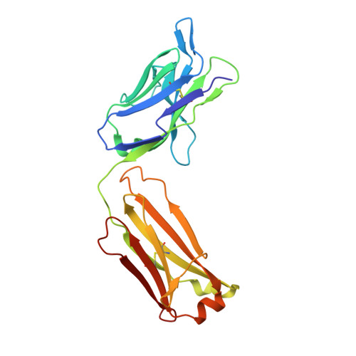
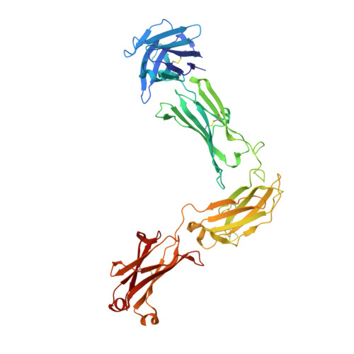
Structure of full-length human anti-PD1 therapeutic IgG4 antibody pembrolizumab.

Scapin, G.Yang, X.Prosise, W.W.McCoy, M.Reichert, P.Johnston, J.M.Kashi, R.S.Strickland, C.

(2015) Nat Struct Mol Biol 22: 953-958

  • DOI: https://doi.org/10.1038/nsmb.3129
  • Primary Citation of Related Structures:  
    5DK3

  • PubMed Abstract: 

    Immunoglobulin G4 antibodies exhibit unusual properties with important biological consequences. We report the structure of the human full-length IgG4 S228P anti-PD1 antibody pembrolizumab, solved to 2.3-Å resolution. Pembrolizumab is a compact molecule, consistent with the presence of a short hinge region. The Fc domain is glycosylated at the CH2 domain on both chains, but one CH2 domain is rotated 120° with respect to the conformation observed in all reported structures to date, and its glycan chain faces the solvent. We speculate that this new conformation is driven by the shorter hinge. The structure suggests a role for the S228P mutation in preventing the IgG4 arm exchange. In addition, this unusual Fc conformation suggests possible structural diversity between IgG subclasses and shows that use of isolated antibody fragments could mask potentially important interactions, owing to molecular flexibility.


  • Organizational Affiliation

    Structural Chemistry, Merck &Co., Inc., Kenilworth, New Jersey, USA.


Macromolecules
Find similar proteins by:  (by identity cutoff)  |  3D Structure
Entity ID: 1
MoleculeChains Sequence LengthOrganismDetailsImage
Light ChainA,
C [auth F]
218Homo sapiensMutation(s): 0 
Entity Groups  
Sequence Clusters30% Identity50% Identity70% Identity90% Identity95% Identity100% Identity
Sequence Annotations
Expand
  • Reference Sequence
Find similar proteins by:  (by identity cutoff)  |  3D Structure
Entity ID: 2
MoleculeChains Sequence LengthOrganismDetailsImage
Heavy ChainB,
D [auth G]
444Homo sapiensMutation(s): 0 
Entity Groups  
Sequence Clusters30% Identity50% Identity70% Identity90% Identity95% Identity100% Identity
Sequence Annotations
Expand
  • Reference Sequence
Oligosaccharides

Help

Entity ID: 3
MoleculeChains Length2D Diagram Glycosylation3D Interactions
2-acetamido-2-deoxy-beta-D-glucopyranose-(1-2)-alpha-D-mannopyranose-(1-3)-[2-acetamido-2-deoxy-beta-D-glucopyranose-(1-2)-alpha-D-mannopyranose-(1-6)]beta-D-mannopyranose-(1-4)-2-acetamido-2-deoxy-beta-D-glucopyranose-(1-4)-2-acetamido-2-deoxy-beta-D-glucopyranoseE [auth C]7N/A
Glycosylation Resources
GlyTouCan:  G39213VZ
GlyCosmos:  G39213VZ
GlyGen:  G39213VZ
Entity ID: 4
MoleculeChains Length2D Diagram Glycosylation3D Interactions
2-acetamido-2-deoxy-beta-D-glucopyranose-(1-2)-alpha-D-mannopyranose-(1-6)-[alpha-D-mannopyranose-(1-3)]beta-D-mannopyranose-(1-4)-2-acetamido-2-deoxy-beta-D-glucopyranose-(1-4)-[alpha-L-fucopyranose-(1-6)]2-acetamido-2-deoxy-beta-D-glucopyranoseF [auth D]7N-Glycosylation
Glycosylation Resources
GlyTouCan:  G45889JQ
GlyCosmos:  G45889JQ
GlyGen:  G45889JQ
Entity ID: 5
MoleculeChains Length2D Diagram Glycosylation3D Interactions
beta-D-fructofuranose-(2-1)-alpha-D-glucopyranoseG [auth E],
H,
I
2N/A
Glycosylation Resources
GlyTouCan:  G05551OP
GlyCosmos:  G05551OP
Biologically Interesting Molecules (External Reference) 1 Unique
Experimental Data & Validation

Experimental Data

  • Method: X-RAY DIFFRACTION
  • Resolution: 2.28 Å
  • R-Value Free: 0.224 
  • R-Value Work: 0.184 
  • R-Value Observed: 0.186 
  • Space Group: P 21 21 21
Unit Cell:
Length ( Å )Angle ( ˚ )
a = 63.83α = 90
b = 110.83β = 90
c = 264.96γ = 90
Software Package:
Software NamePurpose
BUSTERrefinement
Aimlessdata scaling
PDB_EXTRACTdata extraction
XDSdata reduction
PHASERphasing

Structure Validation

View Full Validation Report


Entry History 

Deposition Data

Revision History  (Full details and data files)

  • Version 1.0: 2015-11-18
    Type: Initial release
  • Version 1.1: 2016-07-06
    Changes: Database references
  • Version 1.2: 2016-08-03
    Changes: Database references
  • Version 2.0: 2020-07-29
    Type: Remediation
    Reason: Carbohydrate remediation
    Changes: Advisory, Atomic model, Data collection, Derived calculations, Non-polymer description, Structure summary